Figures & data
Figure 1. All angles were measured from the three reformatted CT slices (coronal, sagittal, and transverse) passing through the center of the femoral head on the ipsilateral side. In the coronal view, the CE angle and the AC angle were measured. The CE angle is defined by the most lateral aspect of the acetabular rim, the center of the femoral head, and a point vertical (superior) to the center point. The AC angle measures the obliqueness of the acetabular roof based on the most lateral and the most medial aspects of the sourcil, and a horizontal (lateral) line. The S-AC angle and the AcetAV are measured from image reformats in the sagittal and transverse planes, respectively. In the sagittal plane, the most anterior portion of the acetabular rim, the top of the acetabular roof, and a horizontal line make the S-AC angle. The acute AcetAV angle is measured by a line parallel to the acetabular opening and one perpendicular to the centers of the femoral heads on the transverse plane. [Color version available online.]
![Figure 1. All angles were measured from the three reformatted CT slices (coronal, sagittal, and transverse) passing through the center of the femoral head on the ipsilateral side. In the coronal view, the CE angle and the AC angle were measured. The CE angle is defined by the most lateral aspect of the acetabular rim, the center of the femoral head, and a point vertical (superior) to the center point. The AC angle measures the obliqueness of the acetabular roof based on the most lateral and the most medial aspects of the sourcil, and a horizontal (lateral) line. The S-AC angle and the AcetAV are measured from image reformats in the sagittal and transverse planes, respectively. In the sagittal plane, the most anterior portion of the acetabular rim, the top of the acetabular roof, and a horizontal line make the S-AC angle. The acute AcetAV angle is measured by a line parallel to the acetabular opening and one perpendicular to the centers of the femoral heads on the transverse plane. [Color version available online.]](/cms/asset/bb3d63df-0f1d-49fe-9689-259ca77e079f/icsu_a_254046_f0001_b.gif)
Figure 2. The oblique slicing method for segmenting the acetabulum lunate. A) Reformatting CT slices are acquired circumferentially about the medial-lateral joint line. This allows the lunate to be traced using the two intersections (medial, lateral) on each slice. B) The creation of the parametric surface implements an oblique plane (blue) rotated about the medial-lateral axis of the joint to find and interpolate points. The acetabulum of this right hip is viewed from the posterior-lateral direction. [Color version available online.]
![Figure 2. The oblique slicing method for segmenting the acetabulum lunate. A) Reformatting CT slices are acquired circumferentially about the medial-lateral joint line. This allows the lunate to be traced using the two intersections (medial, lateral) on each slice. B) The creation of the parametric surface implements an oblique plane (blue) rotated about the medial-lateral axis of the joint to find and interpolate points. The acetabulum of this right hip is viewed from the posterior-lateral direction. [Color version available online.]](/cms/asset/e2985f44-2d1c-4a0f-a47d-26eb1fe10c66/icsu_a_254046_f0002_b.gif)
Figure 3. Transparent views of the acetabulum. The 3D Lunate-Trace curve is shown as a blue outline, and the region in red is the interpolated surface of the acetabulum lunate. The angles are defined using the segmented model, and are analogous to those in . [Color version available online.]
![Figure 3. Transparent views of the acetabulum. The 3D Lunate-Trace curve is shown as a blue outline, and the region in red is the interpolated surface of the acetabulum lunate. The angles are defined using the segmented model, and are analogous to those in Figure 1. [Color version available online.]](/cms/asset/2c9d1669-4ae4-4254-bc8b-a9b78f57ab6c/icsu_a_254046_f0003_b.gif)
Table I. Recorded radiological angle measurements (in degrees) for manual observers (O1–O3) and the computerized Lunate-Trace method (LT) for a total of 24 patients (Pt). A (−) entry indicates that no measurement was recorded because the required anatomical features were not visible on the slice plane.
Figure 4. Histograms of measuring error for observers 1–3 (O1-O3) and the computer assisted Lunate-Trace method (LT).
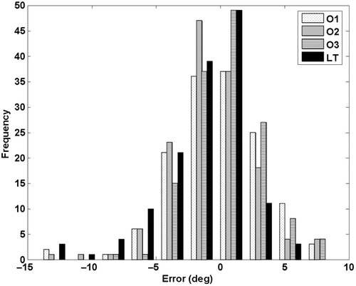
Table II. Error distributions (mean ± standard deviation) for each observer based on the type of angle measured. Two manual observers showed significant (p < 0.01) discrepancies in measuring CE angles. The computerized Lunate-Trace method (LT) showed significant error associated with the S-AC angle measured on the sagittal plane.
Figure 5. Vector plots of location of femoral head with respect to acetabular joint center (mm) in the superior-lateral (frontal plane) and superior-anterior (sagittal plane) directions. [Color version available online.]
![Figure 5. Vector plots of location of femoral head with respect to acetabular joint center (mm) in the superior-lateral (frontal plane) and superior-anterior (sagittal plane) directions. [Color version available online.]](/cms/asset/d111b8aa-5379-4e5f-a801-60b2c5ac5cea/icsu_a_254046_f0005_b.gif)