Figures & data
Figure 1. Anteroposterior (a) and lateral (b) views of a plastic pelvic model with aluminum screws inserted in predetermined anatomical points around the pelvis and acetabulum.
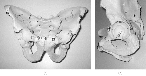
Figure 2. Dial caliper used for manual measurements of point-to-point distances.
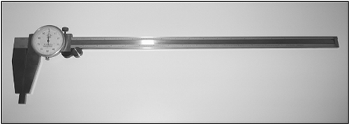
Figure 3. Anthropometric analysis view generated in Mimics software package with screw heads segmented in red and pelvis segmented in green. [Color version available online.]
![Figure 3. Anthropometric analysis view generated in Mimics software package with screw heads segmented in red and pelvis segmented in green. [Color version available online.]](/cms/asset/eb2d0293-5d00-42ea-8901-97ae870d8bbf/icsu_a_267882_f0003_b.gif)
Figure 4. Polygonal structure generated in CAD software package and imported into Mimics with subsequent validation of interplane angular measurements. [Color version available online.]
![Figure 4. Polygonal structure generated in CAD software package and imported into Mimics with subsequent validation of interplane angular measurements. [Color version available online.]](/cms/asset/5cd9cb02-dea5-45fb-9161-1e6caafe48c1/icsu_a_267882_f0004_b.gif)
Figure 5. Two-way ANOVA table for linear displacement with effect of observer and measurement technique (software versus manual) (error bars = 95% confidence interval).
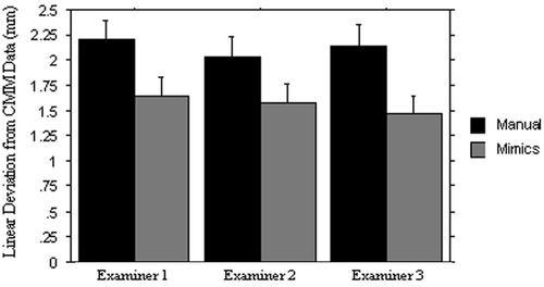
Figure 6. Two-way ANOVA table for angular displacement with effect of observer and measurement technique (software versus manual) (error bars = 95% confidence interval).
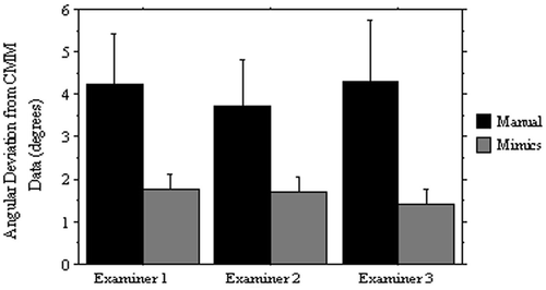
Table I. Intra-class correlation coefficients for manual and software measurements of linear distances and linear angles.
Table II. Expected and measured interplane face angles of polygons imported into the Mimics software package.
Figure 7. Clinical application of the validated interplane measurement protocol to determine variations in acetabular anteversion in a 3D CT-generated computer model of the pelvis. (a) Low magnification view. (b) High magnification view of right acetabulum demonstrating four anteversion planes as measured at different vertical positions of the acetabulum. The superior aspect of the acetabulum is divided into four vertical levels with the lowest level as the equator. Acetabular version at level 1 (green, long black arrow) shows retroversion of 7.75°; level 2 (white, small black arrowhead), level 3 (yellow, large white arrowhead), and level 4 or the equator (red, large white arrow) all demonstrate anteversion of the acetabulum of 19.47°, 21.52°, and 13.55°, respectively. [Color version available online.]
![Figure 7. Clinical application of the validated interplane measurement protocol to determine variations in acetabular anteversion in a 3D CT-generated computer model of the pelvis. (a) Low magnification view. (b) High magnification view of right acetabulum demonstrating four anteversion planes as measured at different vertical positions of the acetabulum. The superior aspect of the acetabulum is divided into four vertical levels with the lowest level as the equator. Acetabular version at level 1 (green, long black arrow) shows retroversion of 7.75°; level 2 (white, small black arrowhead), level 3 (yellow, large white arrowhead), and level 4 or the equator (red, large white arrow) all demonstrate anteversion of the acetabulum of 19.47°, 21.52°, and 13.55°, respectively. [Color version available online.]](/cms/asset/1163224a-ad32-4972-be64-e72b201ad68b/icsu_a_267882_f0007_b.gif)
Figure 8. Presurgical planning and performance of a Bernese periacetabular osteotomy on a 3D model. This technique permits preoperative optimization of anterior and lateral acetabular roof coverage, and medialization with detection of bone interference. [Color version available online.]
![Figure 8. Presurgical planning and performance of a Bernese periacetabular osteotomy on a 3D model. This technique permits preoperative optimization of anterior and lateral acetabular roof coverage, and medialization with detection of bone interference. [Color version available online.]](/cms/asset/5b3db2df-bbb7-484f-8de6-40306edaa314/icsu_a_267882_f0008_b.gif)