Figures & data
Figure 1. Endoscopic view of a transgastric procedure. [Color version available online.]
![Figure 1. Endoscopic view of a transgastric procedure. [Color version available online.]](/cms/asset/07cc2a42-afac-4311-beb6-9fd303cae7d6/icsu_a_274646_f0001_b.gif)
Figure 2. System description: data flow paths and main displays. [Color version available online.]
![Figure 2. System description: data flow paths and main displays. [Color version available online.]](/cms/asset/22f62a1b-8ff1-49bf-ba77-1d1fb8c0d4c5/icsu_a_274646_f0002_b.gif)
Figure 3. View of the system interface as presented to the clinician. [Color version available online.]
![Figure 3. View of the system interface as presented to the clinician. [Color version available online.]](/cms/asset/8d499789-81e6-45c1-b884-af77ba779154/icsu_a_274646_f0003_b.gif)
Figure 4. The sensor attached to the endoscope tip.
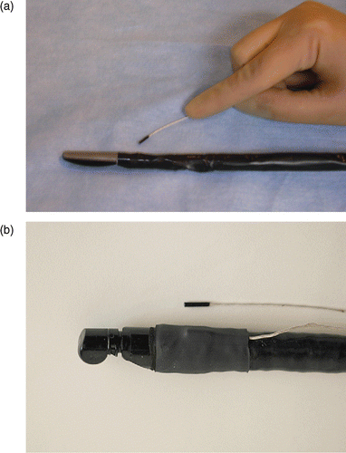
Figure 5. Landmark points chosen for system registration error validation and corresponding examples of the Doppler US views for each landmark point. The branching interface is coded in the Doppler US as a change of color due to a change in blood flow direction. [Color version available online.]
![Figure 5. Landmark points chosen for system registration error validation and corresponding examples of the Doppler US views for each landmark point. The branching interface is coded in the Doppler US as a change of color due to a change in blood flow direction. [Color version available online.]](/cms/asset/cb6335da-4580-4a6a-a227-4cb4397b5596/icsu_a_274646_f0005_b.gif)
Figure 6. Registration error definitions.
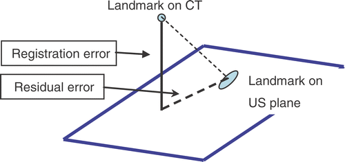
Figure 7. Registration error measurements using box-and-whisker plots. (a) Error for each case considering the landmarks altogether. (b) Error for each landmark considering the cases altogether. [Color version available online.]
![Figure 7. Registration error measurements using box-and-whisker plots. (a) Error for each case considering the landmarks altogether. (b) Error for each landmark considering the cases altogether. [Color version available online.]](/cms/asset/ba78254e-3007-401e-b1e8-1f87f8a30ec4/icsu_a_274646_f0007_b.gif)
Figure 8. Histogram of the total registration error considering all the landmarks and all the cases in our sample population.
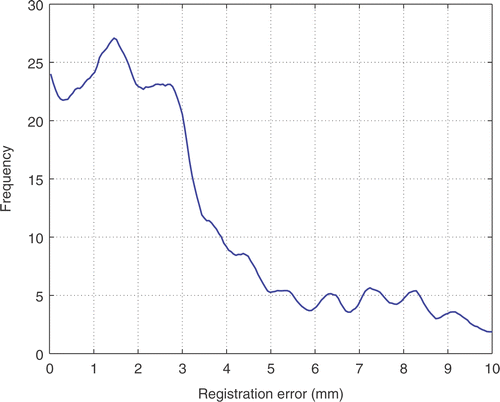
Table I. Total registration error statistical analysis. Left: Bootstrap estimates for the mean, median and standard deviation (SD) of the out-of-plane error. Right: Bootstrap estimates for the cumulative probability of the out-of-plane error at 5 mm, 7 mm and 8 mm.
Figure 9. Two examples of system performance during the evaluation process. The images show how the contextual information added by the three displays improves the awareness of the operator regarding the structure being imaged by the US. [Color version available online.]
![Figure 9. Two examples of system performance during the evaluation process. The images show how the contextual information added by the three displays improves the awareness of the operator regarding the structure being imaged by the US. [Color version available online.]](/cms/asset/14255342-f79d-43d4-b3e2-4ad4ecf3c17e/icsu_a_274646_f0009_b.gif)
Figure 10. Spectrum of the time series corresponding to forced breathing. A harmonic at approximately 0.6 Hz represents the main breathing component.
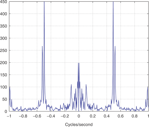
Table II. Kinematic analysis comparing our system (IRGUS) and the conventional endoscopic approach (EUS). See reference 25 for a description of the parameters and analysis process.
Figure 11. Challenges for optimal stomach access to the abdominal cavity. (a) View of the vasculature anatomy of the stomach in a porcine model. The right image is an exterior view of the right portion of the specimen. The right gastrodoudenal artery (white arrow) can be seen lying on top of the stomach wall. The left image is an endoscopic view of the same site from inside the stomach looking towards the right-anterior part. The vessel structure has been overlaid with the semi-transparent model of the stomach wall, but this will not be visible in a standard endoscopic view. (b) A distance map to stomach vessels on the stomach surface. [Color version available online.]
![Figure 11. Challenges for optimal stomach access to the abdominal cavity. (a) View of the vasculature anatomy of the stomach in a porcine model. The right image is an exterior view of the right portion of the specimen. The right gastrodoudenal artery (white arrow) can be seen lying on top of the stomach wall. The left image is an endoscopic view of the same site from inside the stomach looking towards the right-anterior part. The vessel structure has been overlaid with the semi-transparent model of the stomach wall, but this will not be visible in a standard endoscopic view. (b) A distance map to stomach vessels on the stomach surface. [Color version available online.]](/cms/asset/92db592d-4e13-46f9-9a23-b9c9e3bc7b64/icsu_a_274646_f0011_b.gif)