Figures & data
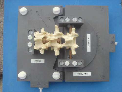
Figure 1. The phantom. Three vertebrae are attached to a base plate. L4 and L5 are secured rigidly to the base plate, and L3 is translated using screws.

Table I. Plastic and human vertebrae in different 3D translation positions in the phantom.
Figure 2. (a) Simultaneous display of two CT volumes (reference [left] and target [right]) in axial, coronal and sagittal views. A single landmark in the L4 verterbra in each volume is shown (blue diamond) on the orthogonal slices that intersect it. Co-homologous points are designated in both volumes and used to bring the L4 in the target volume into spatial alignment with the L4 in the reference volume. For this study, nine landmarks in L4 where used. (b) Close-up coronal view of human vertebra with co-homologous landmarks shown (orange diamonds) in the reference (top) and target (bottom) volumes. [Color version available online.]
![Figure 2. (a) Simultaneous display of two CT volumes (reference [left] and target [right]) in axial, coronal and sagittal views. A single landmark in the L4 verterbra in each volume is shown (blue diamond) on the orthogonal slices that intersect it. Co-homologous points are designated in both volumes and used to bring the L4 in the target volume into spatial alignment with the L4 in the reference volume. For this study, nine landmarks in L4 where used. (b) Close-up coronal view of human vertebra with co-homologous landmarks shown (orange diamonds) in the reference (top) and target (bottom) volumes. [Color version available online.]](/cms/asset/7660d64d-0f7f-4acd-8116-18187433429b/icsu_a_288423_f0002_b.gif)
Figure 3. (a) A 3D display of L4 after registration (case 34). Note the overlapping “zebra-like” pattern between the reference volume isosurface (yellow) and the transformed volume isosurface (green) created when the two surfaces coincide. This pattern indicates that the registration is better than the smallest image element (voxel) in the volumes. (b) A 2D overlay axial view where the gray (left) side of the vertebra is the reference volume and the red (right) side is an overlay of the transformed volume on the reference volume. Note the close match between all the condensed areas of the vertebra (case 34). [Color version available online.]
![Figure 3. (a) A 3D display of L4 after registration (case 34). Note the overlapping “zebra-like” pattern between the reference volume isosurface (yellow) and the transformed volume isosurface (green) created when the two surfaces coincide. This pattern indicates that the registration is better than the smallest image element (voxel) in the volumes. (b) A 2D overlay axial view where the gray (left) side of the vertebra is the reference volume and the red (right) side is an overlay of the transformed volume on the reference volume. Note the close match between all the condensed areas of the vertebra (case 34). [Color version available online.]](/cms/asset/152bc40c-fcef-4c23-9ec4-f85c03fc6a37/icsu_a_288423_f0003_b.gif)
Figure 4. Three-dimensional isosurface displays after registration of L4. (a) A case with no movement of L3, showing “zebra” pattern between the reference (yellow) and transformed vertebrae (green) in both L4 and L3. A disc prosthesis is incorporated between the vertebrae for future studies. (b) A 0.5-mm sagittal translation of L5. (c) A 1.0-mm sagittal translation of L3. (d) A 2.0-mm sagittal translation of L3. [Color version available online.]
![Figure 4. Three-dimensional isosurface displays after registration of L4. (a) A case with no movement of L3, showing “zebra” pattern between the reference (yellow) and transformed vertebrae (green) in both L4 and L3. A disc prosthesis is incorporated between the vertebrae for future studies. (b) A 0.5-mm sagittal translation of L5. (c) A 1.0-mm sagittal translation of L3. (d) A 2.0-mm sagittal translation of L3. [Color version available online.]](/cms/asset/3304e997-10c1-40ef-9bce-23ed1a8ba698/icsu_a_288423_f0004_b.gif)
Table II. Repeatability. Difference (in mm) when 10 cases where analyzed twice under repeatability conditions.