Figures & data
Figure 1. The Herbert bone screw.
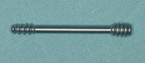
Figure 2. Phantom model of the lower jaw with a radiologically detectable copper wire representing the inferior alveolar nerve and an artificial mandibular angle fracture. [Color version available online.]
![Figure 2. Phantom model of the lower jaw with a radiologically detectable copper wire representing the inferior alveolar nerve and an artificial mandibular angle fracture. [Color version available online.]](/cms/asset/02f88ab8-0024-4990-880f-37523d3d99a2/icsu_a_288426_f0002_b.gif)
Figure 3. X-ray of the phantom model. Four titanium screws in the alveolar bone act as reference markers.
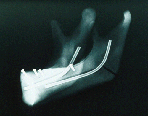
Figure 4. The approach and drilling depth are planned on the workstation monitor. [Color version available online.]
![Figure 4. The approach and drilling depth are planned on the workstation monitor. [Color version available online.]](/cms/asset/4784caa6-2831-4960-bd2e-981310082149/icsu_a_288426_f0004_b.gif)
Figure 5. The drill wire is calibrated in the calibration station. A trackable reference frame is fixed on the drilling handle.
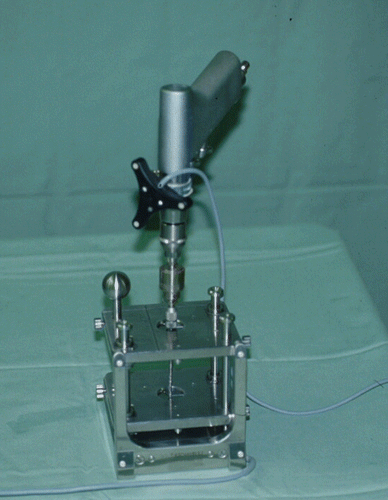
Figure 6. Insertion of the drill wire with a fixed mandibular model and a trackable reference frame.
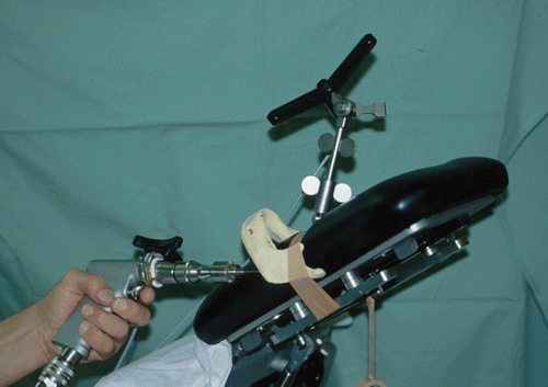
Figure 7. Corresponding view of the workstation monitor showing the preoperatively planned drilling path (yellow line) and the actual path of the drill wire (green line); the coronal layer with parallel follow-up along the preoperatively planned drilling pathway to the target (upper left); the sagittal layer (upper right); the axial layer (lower left); and the 3D depiction (lower right). [Color version available online.]
![Figure 7. Corresponding view of the workstation monitor showing the preoperatively planned drilling path (yellow line) and the actual path of the drill wire (green line); the coronal layer with parallel follow-up along the preoperatively planned drilling pathway to the target (upper left); the sagittal layer (upper right); the axial layer (lower left); and the 3D depiction (lower right). [Color version available online.]](/cms/asset/614a64d4-cada-4281-86f6-0b99c42e0f3b/icsu_a_288426_f0007_b.gif)
Figure 8. Depiction aid with superimposition of the inner and outer crosshairs on the workstation. [Color version available online.]
![Figure 8. Depiction aid with superimposition of the inner and outer crosshairs on the workstation. [Color version available online.]](/cms/asset/9cca94c5-67cc-4044-8b5a-180182f9de31/icsu_a_288426_f0008_b.gif)
Figure 9. Mandibular model with the inserted bone screw. A perforation of the outer cortical layer can be seen. [Color version available online.]
![Figure 9. Mandibular model with the inserted bone screw. A perforation of the outer cortical layer can be seen. [Color version available online.]](/cms/asset/9ce016a2-954f-4785-9b4c-4ce7b8fe302a/icsu_a_288426_f0009_b.gif)
Figure 10. Postoperative X-ray of the mandibular model with inserted screw. [Color version available online.]
![Figure 10. Postoperative X-ray of the mandibular model with inserted screw. [Color version available online.]](/cms/asset/6f7c671a-e386-4178-a51b-228adbcabcdc/icsu_a_288426_f0010_b.gif)
Table I. Results of ten target drillings in phantom models of the lower jaw. The mean planned insertion depth on the computer monitor was 0.17 mm longer than the mean actual depth measured in the phantom models.