Figures & data
Table I. CT surface mesh classes, using the self-developed Filter & Mesh procedure of Gelaude et al. Citation[25]. (pps: points per spline; spAcc: spline accuracy value; IsDist: interslice distance. Each class consists of 15 entire femur meshes; notice that a smaller spline accuracy value (spAcc) designates a higher spline accuracy.) Note: from this table, “groups” of classes were defined to investigate the influence of one parameter setting when all others were kept unchanged, e.g., groups 215, 316 and 347, which check the influence of a varying IsDist, spAcc and pps, respectively.
Table II. CT surface mesh classes, using Mimics® (Version 10.0). (resolution = with regard to matrix reduction, how many voxels were geometrically merged and averaged by their intensity value; * 2 iterations, smoothing factor 0.3; ** advance edge reduction algorithm, tolerance (pixel size)/8, edge angle 10, iteration 3. All classes use the contour interpolation method (2D partial volume compensation) and an interslice distance of 1 mm; each class consists of 15 entire femur meshes).
Figure 1. Experimental set-up for optical surface scanning with the Metris LC50, mounted on a Coord3 MC16 coordinate measuring machine. The dry femur is mounted by inserting a rigid pin distally in the intramedullary canal, and is positioned obliquely with respect to the ground plane. Pre-drilling of the femur with a slightly smaller diameter than the pin diameter ensures rotational stability of the bone.
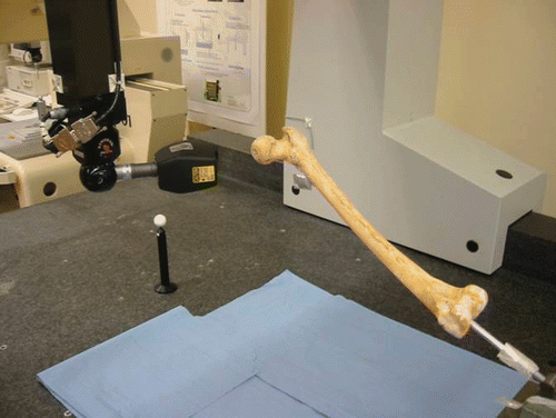
Figure 2. Comparison scheme. To assess the combined influence of soft tissue and boiling on the comparisons “A” between dry bone ground truths and bone meshes segmented from intact cadaver CT scans, additional dry bone CT scans are similarly processed (“B”) by one of the class L, m or h segmentation algorithms (). Relative comparison of the 3D deviation statistics (mean X, standard deviation σ) for A and B then cancels out the contribution of the segmentation algorithm, and–if the same scanner and settings are used–of the CT machine as well.
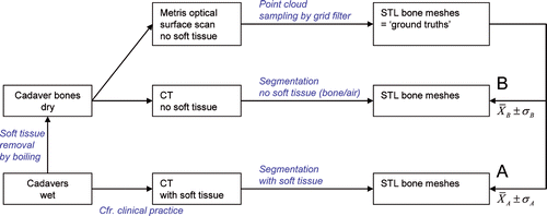
Figure 3. Graphs showing 3D deviations of CT meshes for the entire femur. (pps = points per spline; spAcc = spline accuracy value; IsDist = interslice distance. Note that a smaller spline accuracy value (spAcc) designates a higher spline accuracy.) (See and for class ID and for statistics.) [Color version available online.]
![Figure 3. Graphs showing 3D deviations of CT meshes for the entire femur. (pps = points per spline; spAcc = spline accuracy value; IsDist = interslice distance. Note that a smaller spline accuracy value (spAcc) designates a higher spline accuracy.) (See Tables I and II for class ID and Table III for statistics.) [Color version available online.]](/cms/asset/bcda2d5d-cfe7-426b-8aaf-d8c37a19dbd4/icsu_a_319745_f0003_b.gif)
Table III. Three-dimensional deviations of cadaver CT femur meshes (in mm). (X: mean; σ: standard deviation; A: see ; class ID: see and ; n: number of measurements; a negative 3D deviation indicates that the CT mesh is inflated.)
Table IV. Three-dimensional deviations of dry bone CT meshes (in mm) using segmentation algorithm of class h (see ) (X: mean; σ: standard deviation; n: number of measurements; a negative 3D deviation indicates that the CT mesh is inflated. For “A” and “B”, see ).
Figure 4. Influence of soft tissue removal and boiling of the cadaver bones. (a) For class h (see and the section headed Assessing influence of soft tissue presence and boiling), the dry bone CT scans (B) as well as the intact specimen CT scans (A) of all specimens are segmented and compared to the MetrisScan ground truths (entire femora). The mean 3D deviation XB is attributed to the CT machine and segmentation algorithm; apart from the same CT machine and segmentation algorithm, the mean 3D deviation XA is attributed to soft tissue presence (setting the bone Hounsfield window for cortical bone) and boiling of the cadaver bones. (b) Graphs of the 3D deviations for all intact specimen CT-based meshes, all classes (entire femora). A positive shift Δ XAB should be applied to the graphs to counteract the combined effect of soft tissue presence and boiling. The dotted vertical line should serve as new zero line. [Color version available online.]
![Figure 4. Influence of soft tissue removal and boiling of the cadaver bones. (a) For class h (see Table II and the section headed Assessing influence of soft tissue presence and boiling), the dry bone CT scans (B) as well as the intact specimen CT scans (A) of all specimens are segmented and compared to the MetrisScan ground truths (entire femora). The mean 3D deviation XB is attributed to the CT machine and segmentation algorithm; apart from the same CT machine and segmentation algorithm, the mean 3D deviation XA is attributed to soft tissue presence (setting the bone Hounsfield window for cortical bone) and boiling of the cadaver bones. (b) Graphs of the 3D deviations for all intact specimen CT-based meshes, all classes (entire femora). A positive shift Δ XAB should be applied to the graphs to counteract the combined effect of soft tissue presence and boiling. The dotted vertical line should serve as new zero line. [Color version available online.]](/cms/asset/f6d44c59-8d68-4624-a017-c711ac59e391/icsu_a_319745_f0004_b.gif)
Table V. Optimal choice of segmentation parameters for outer surface mesh generation for femoral CT scans with soft tissue, as in clinical practice. (See and for class definitions; voxel in-slice dimension = 1 mm × 1 mm; a negative 3D deviation indicates that the CT mesh is inflated; Δ XAB = compensation for combined effect of soft tissue presence during bone Hounsfield windowing and shrinkage of the ground truth femora by boiling [see ].)