Figures & data
Figure 1. Navigation system for targeted prostate biopsy. (a) System components: localizer (L), tracked ultrasound probe (US) and prostate phantom (P). (b) System setup in patient study. [Color version available online.]
![Figure 1. Navigation system for targeted prostate biopsy. (a) System components: localizer (L), tracked ultrasound probe (US) and prostate phantom (P). (b) System setup in patient study. [Color version available online.]](/cms/asset/85a3e89f-5dcc-4596-8c35-abae20a1f092/icsu_a_336631_f0001_b.gif)
Figure 2. Selected image frames for 2.5D to 3D registration. [Color version available online.]
![Figure 2. Selected image frames for 2.5D to 3D registration. [Color version available online.]](/cms/asset/9c364f69-812d-42fc-b305-4a745ee2fd3d/icsu_a_336631_f0002_b.gif)
Figure 3. Example of image-based motion compensation. (a) Real-time ultrasound. (b) Registration result of (a) in the reconstructed reference ultrasound volume. (c) Initial starting point of the registration.
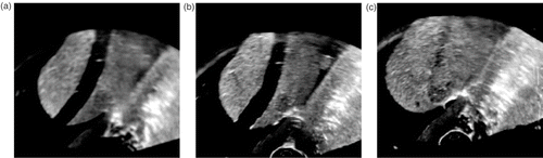
Figure 4. CT image example showing needle tip in tumor. (a) XY section. (b) ZY section. (c) XZ section. [Color version available online.]
![Figure 4. CT image example showing needle tip in tumor. (a) XY section. (b) ZY section. (c) XZ section. [Color version available online.]](/cms/asset/8c783081-63be-493a-ad98-4ab30b7a074f/icsu_a_336631_f0004_b.gif)
Figure 5. 2D plots of the objective function near the global minimum with respect to two translation parameters. (a) is the result of registering one image frame, while (b) is the result of registering four image frames. The grid unit is 1 mm. [Color version available online.]
![Figure 5. 2D plots of the objective function near the global minimum with respect to two translation parameters. (a) is the result of registering one image frame, while (b) is the result of registering four image frames. The grid unit is 1 mm. [Color version available online.]](/cms/asset/487b2dce-48be-4793-ac3a-bf97e7dd06fe/icsu_a_336631_f0005_b.gif)
Figure 6. Motion compensation using 2.5D/3D registration. The red contours show the prostate segmentation in the MRI image. The 3D MRI volume is pre-registered to a 3D ultrasound volume that is not shown. Top row: RTUS overlaid on MRI. Bottom row: MRI images. (a) and (a′) are the initial registration without patient motion; (b) and (b′) are the deteriorated registration after patient motion; and (c) and (c′) are the registration after motion compensation. [Color version available online.]
![Figure 6. Motion compensation using 2.5D/3D registration. The red contours show the prostate segmentation in the MRI image. The 3D MRI volume is pre-registered to a 3D ultrasound volume that is not shown. Top row: RTUS overlaid on MRI. Bottom row: MRI images. (a) and (a′) are the initial registration without patient motion; (b) and (b′) are the deteriorated registration after patient motion; and (c) and (c′) are the registration after motion compensation. [Color version available online.]](/cms/asset/89981e40-5ff2-4c98-8cd6-cf6e7b1a98bb/icsu_a_336631_f0006_b.gif)
Figure 7. Screenshots of fused MRI/TRUS image guidance. (a) T2-weighted MRI fused with reference ultrasound volume (color map), along with the real-time ultrasound and target information. (b) Corresponding real-time ultrasound (top) and MRI MPR views (bottom) at the time of needle deployment. [Color version available online.]
![Figure 7. Screenshots of fused MRI/TRUS image guidance. (a) T2-weighted MRI fused with reference ultrasound volume (color map), along with the real-time ultrasound and target information. (b) Corresponding real-time ultrasound (top) and MRI MPR views (bottom) at the time of needle deployment. [Color version available online.]](/cms/asset/61758e2e-b5ca-4a55-bd9e-dd63df6e99fe/icsu_a_336631_f0007_b.gif)