Figures & data
Figure 1. Schematic illustration demonstrating a surface NIR light emitter coupled to a low-light-sensitive detector. The cortical hemodynamic change from a banana-shaped volume of tissue between the emitter and detector is probed non-invasively. Application of a series of emitters and detectors results in hemodynamic recordings from multiple cortical sites (channels) simultaneously.
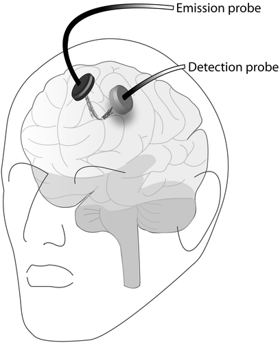
Figure 2. The experimental set-up. Schematic illustration of a study subject engaged in a knot-tying drill. The electromagnetic pulse emitter is shown on the table in front of the knot-tying jig and motion-tracking sensors are visible on the dorsum of the subject's hands. The figure illustrates the 3 × 3 arrangement of NIRS optodes and the relative positions of NIR emitters (red) and detectors (blue). Optode location is obtained by transferring topographic data from a representative subject to a 3D cortical surface image of a high-resolution T1-weighted MRI image. The two subplots illustrate the location of NIR channels (shaded circles) on the cortical surfaces. The approximate International 10-10 landmarks are highlighted (yellow boxes). [Color version available online.]
![Figure 2. The experimental set-up. Schematic illustration of a study subject engaged in a knot-tying drill. The electromagnetic pulse emitter is shown on the table in front of the knot-tying jig and motion-tracking sensors are visible on the dorsum of the subject's hands. The figure illustrates the 3 × 3 arrangement of NIRS optodes and the relative positions of NIR emitters (red) and detectors (blue). Optode location is obtained by transferring topographic data from a representative subject to a 3D cortical surface image of a high-resolution T1-weighted MRI image. The two subplots illustrate the location of NIR channels (shaded circles) on the cortical surfaces. The approximate International 10-10 landmarks are highlighted (yellow boxes). [Color version available online.]](/cms/asset/1bd81441-1ef9-4eda-8075-d212429c9180/icsu_a_353316_f0002_b.gif)
Figure 3. Schematic illustration demonstrating the application and flow of the FR-Isomap data embedding technique.
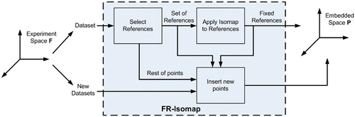
Table I. Comparison of knot-tying performance according to expertise. Data are presented as median [range]. Significant p-values ≤ 0.001 are in bold.
Table II. Comparison of knot-tying performance in medical students before and after a week of practice. Data are presented as median [range]. Significant p-values ≤ 0.001 (Wilcoxon Sign Rank test) are in bold.
Figure 4. (a) Graph depicting residual variance of the fixed references training set in FR-Isomap for fNIRS data from expert and novice surgeons. (b) Diagram of the manifold embedding results for study (A), illustrating the distribution of channel responses and how it varies along the first two principal dimensions for all subjects studied. Locations of the original signals (a–f) are marked on the embedded space, demonstrating the intrinsic trend captured by the embedding technique. (Originally published in reference Citation[12]. Reprinted with permission from Springer Science and Business Media.) (c) The intensity plot of change in total hemoglobin (HbT) demonstrates regions of the embedding most likely to represent brain activation. [Color version available online.]
![Figure 4. (a) Graph depicting residual variance of the fixed references training set in FR-Isomap for fNIRS data from expert and novice surgeons. (b) Diagram of the manifold embedding results for study (A), illustrating the distribution of channel responses and how it varies along the first two principal dimensions for all subjects studied. Locations of the original signals (a–f) are marked on the embedded space, demonstrating the intrinsic trend captured by the embedding technique. (Originally published in reference Citation[12]. Reprinted with permission from Springer Science and Business Media.) (c) The intensity plot of change in total hemoglobin (HbT) demonstrates regions of the embedding most likely to represent brain activation. [Color version available online.]](/cms/asset/13d6a4bb-577c-4d25-8ed9-9b3024d0dbe5/icsu_a_353316_f0004_b.gif)
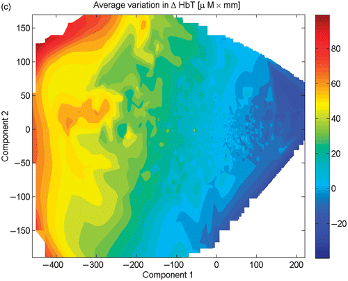
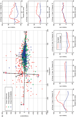
Figure 5. Manifold embedding results depicting practice-dependent reorganization of cortical behavior in surgical novices. Data represents 74-D trial HbO2 and HHb values for a given channel and subject. Trajectories illustrate the transition between first-session data (pink crosses) and second-session data (green circles).
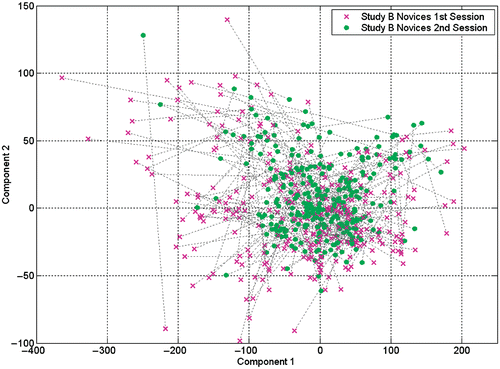
Figure 6. Using the k-means algorithm, the central point (centroid) for each group was located in the embedded space. The average distance between each centroid and all other points for that group was calculated (colored spheres), and the distance to the farthest point (cross) is shown as a solid line. [Color version available online.]
![Figure 6. Using the k-means algorithm, the central point (centroid) for each group was located in the embedded space. The average distance between each centroid and all other points for that group was calculated (colored spheres), and the distance to the farthest point (cross) is shown as a solid line. [Color version available online.]](/cms/asset/76007bf9-d1bd-496d-9975-e291d4d936c1/icsu_a_353316_f0006_b.gif)
Figure 7. (a) Between-group Earth Mover's Distance (EMD) results for study (A); and (b) comparison between studies (A) and (B). [Color version available online.]
![Figure 7. (a) Between-group Earth Mover's Distance (EMD) results for study (A); and (b) comparison between studies (A) and (B). [Color version available online.]](/cms/asset/fa6e34d9-2808-4389-a75f-67a8e83de562/icsu_a_353316_f0007_b.gif)
Table III. Results of a random effect model for learning-related changes in (a) ΔHbO2, (b) ΔHHb and (c) ΔHbT in surgical novices. s.e = standard error. Significant p-values < 0.01 are in bold, and significant values < 0.001 are in bold italics.