Figures & data
Figure 1. Definitions of the twelve projection parameters. Six parameters (i.e., 3 translations and 3 angulations) are required to quantify the fracture deformity and a further six parameters (i.e., 3 translations and 3 angulations) are required to quantify the spatial relationship between the proximal pin clamp and the proximal fracture fragment. (1) Angulation parameters: Lines are drawn along the longitudinal axes of the proximal and distal fracture fragments on both the AP (A) and lateral (B) X-ray images. The angles between them are the AP view fracture site angulation (αf) and the lateral view fracture site angulation (βf), respectively. Lines are also drawn along the longitudinal edge of the proximal pin clamp on the AP and lateral X-ray images. The angles between the pin clamp line and the proximal fracture fragment line on the AP and lateral view are the AP view mounting angulation (αm) and lateral view mounting angulation (βm), respectively. On the transverse projection photograph (C), the angle between the AP axes of the distal and proximal fracture fragments is the transverse view fracture site angulation (γf). The angle between a line along the distal bone pin of the proximal pin clamp and the lateral axis of the proximal fracture fragment is the rotational mounting offset (γm). With the frontal plane of the proximal tibial fragment positioned parallel to an examination table, the AP and lateral axes of the proximal fracture fragment correspond to vertical and horizontal lines on the transverse photograph. (2) Translation parameters: On both the AP (A) and lateral (B) X-ray images, the origins of each fracture fragment are defined as the centroid of the outline of each fracture end. The three components of fracture site translations (xf, yf, zf) correspond to the x, y, z components of a vector from the origin of the proximal fracture fragment to the origin of the distal fracture fragment in terms of the local coordinate system of the proximal fracture fragment. The origin of the proximal pin clamp is defined as the tip of the distal bone pin of the proximal pin clamp. Hence, the three components of mounting translations (xm, ym, zm) are the x, y, z components of a vector from the origin of the proximal fracture fragment to the origin of the proximal pin clamp in terms of the local coordinate system of the proximal fracture fragment. zf and zm can be measured in both the AP and lateral view.
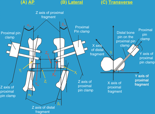
Figure 2. Workflow for using the Bone Reposition Device (BRD) as a template to guide the reduction process (1 = pin offset locator).
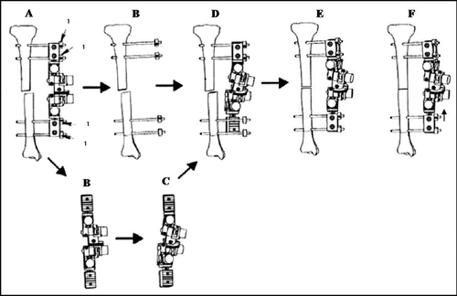
Figure 3. Anteroposterior (AP) X-ray (A), lateral X-ray (B) and transverse photo images (C) of a tibia phantom used in the laboratory validation. Lines and origins were superimposed and six deformity and six mounting parameters were then measured from the images. Pin offset distance and 7 joint configuration parameters were also measured preoperatively.
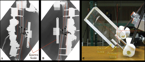
Table I. Initial deformities of the five displaced tibial fracture models.
Figure 4. Bone alignment before and after projection-based fracture reduction of model A. Left: Before reduction (θr = 22.76°, dr = 29.28 mm); Right: After reduction (θr = 3.07°, dr = 1.41 mm).
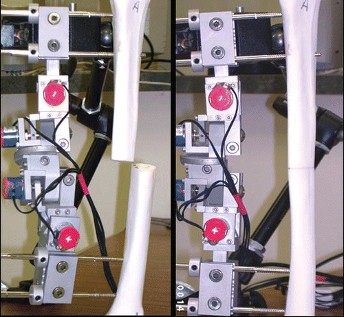
Table II. Reduction accuracy for the five tibia phantoms.
Table III. Inter-rater reliability of the projection parameters on the X-ray images.
Figure 5. Effects of limb positioning errors on angulation parameters: Measured changes in angulation parameters (αf, αm, βf, βm, γf, γm). Changes are expressed as the mean absolute difference between the parameter as measured in the true frontal, sagittal and transverse planes and the same parameter as measured in a plane with introduced error of ±10° about the axis indicated.
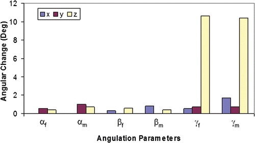
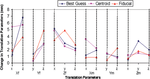
Figure 6. Effects of limb positioning errors on translation parameters: Measured changes in translation parameters (Xf, Yf, Zf, Xm, Ym, Zm). Changes are expressed as the mean absolute difference between the parameter as measured in the true frontal, sagittal and transverse planes and the same parameter as measured in a plane with introduced error (x, y, z indicate the axis about which ±10° was introduced). The parameters resulting from three methods of identifying the fragment origins (Best Guess, Centroid, and Fiducial) are compared.