713
Views
11
CrossRef citations to date
0
Altmetric
Research Article

Navigation Endoscopic Assisted Tumor (NEAT) surgery for benign bone tumors of the extremities

, , , &
Pages 32-39 | Received 16 Jul 2009, Accepted 08 Mar 2010, Published online: 30 Apr 2010

Figures & data

Table I.  Details of five cases with navigation endoscopic assisted intra-lesional curettage.

Figure 1. CT and MR images for Case 2 were fused in the navigation system and the tumor delineated from the fused image datasets. The tumor volume (outlined in red) can be calculated, enabling an estimate of the amount of bone cement or artificial bone graft required to fill the bone defect after intra-lesional curettage. The locations of working portals were planned and marked by virtual pedicle screws.
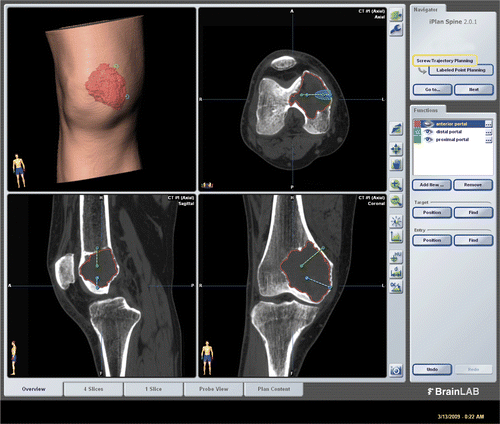
Figure 1. CT and MR images for Case 2 were fused in the navigation system and the tumor delineated from the fused image datasets. The tumor volume (outlined in red) can be calculated, enabling an estimate of the amount of bone cement or artificial bone graft required to fill the bone defect after intra-lesional curettage. The locations of working portals were planned and marked by virtual pedicle screws.

Figure 2. The set-up for NEAT surgery. A shoulder arthroscope and high-speed bone burr mounting a navigation tracker were inserted via two working portals into the tumor cavity. Intra-lesional curettage could be performed under both direct endoscopic visualization and navigation guidance.
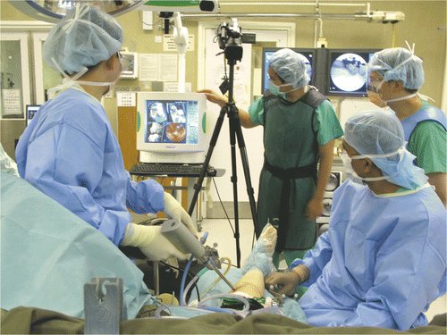
Figure 2. The set-up for NEAT surgery. A shoulder arthroscope and high-speed bone burr mounting a navigation tracker were inserted via two working portals into the tumor cavity. Intra-lesional curettage could be performed under both direct endoscopic visualization and navigation guidance.

Figure 3. CT-fluoro matching was used for image-to-patient registration in Case 1. This involved the matching of preoperative CT images of a segmented bone region with two intraoperatively acquired fluoroscopic images, thus allowing minimally access registration of the preoperative CT images.
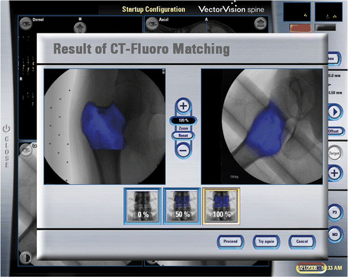
Figure 3. CT-fluoro matching was used for image-to-patient registration in Case 1. This involved the matching of preoperative CT images of a segmented bone region with two intraoperatively acquired fluoroscopic images, thus allowing minimally access registration of the preoperative CT images.

Figure 4. The tumor cavity and bone burring procedure could be directly visualized with the endoscope via small incision portal sites. The location of the navigated instrument tip (the orange circle) is shown with respect to reconstructed CT images. (The tumor is shaded in orange. The extrapolated orange line represents the direction of the instrument and is marked with 5-mm gradations, enabling it to be used to estimate the amount of bone removed and the risk of cortex perforation.)
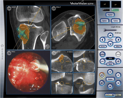
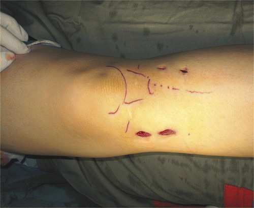
Figure 4. The tumor cavity and bone burring procedure could be directly visualized with the endoscope via small incision portal sites. The location of the navigated instrument tip (the orange circle) is shown with respect to reconstructed CT images. (The tumor is shaded in orange. The extrapolated orange line represents the direction of the instrument and is marked with 5-mm gradations, enabling it to be used to estimate the amount of bone removed and the risk of cortex perforation.)
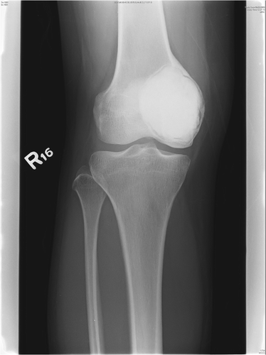
Figure 5. The wounds for Case 3, a patient with a giant cell tumor of the proximal tibia. The two wounds on the medial side served as working portals, while those on the lateral side were used for pin-insertion of the patient tracker.

Figure 5. The wounds for Case 3, a patient with a giant cell tumor of the proximal tibia. The two wounds on the medial side served as working portals, while those on the lateral side were used for pin-insertion of the patient tracker.

Figure 6. Antero-posterior radiograph of the knee of Case 4 at 6 months post-surgery, showing cementation of the bone defect after tumor removal.

Figure 6. Antero-posterior radiograph of the knee of Case 4 at 6 months post-surgery, showing cementation of the bone defect after tumor removal.

Reprints and Corporate Permissions

Please note: Selecting permissions does not provide access to the full text of the article, please see our help page How do I view content?

To request a reprint or corporate permissions for this article, please click on the relevant link below:

Academic Permissions

Please note: Selecting permissions does not provide access to the full text of the article, please see our help page How do I view content?

Obtain permissions instantly via Rightslink by clicking on the button below:

If you are unable to obtain permissions via Rightslink, please complete and submit this Permissions form. For more information, please visit our Permissions help page.