571
Views
8
CrossRef citations to date
0
Altmetric
Clinical Paper

Effect of CT slice thickness on accuracy of implant positioning in navigated total hip arthroplasty

, , , , , & show all
Pages 83-89 | Received 08 Feb 2010, Accepted 10 Sep 2010, Published online: 02 Nov 2010

Figures & data

Figure 1. Screen display showing the inclination and anteversion of the cup, the anteversion and varus/valgus angle of the stem, and the leg length compared with the contralateral side.
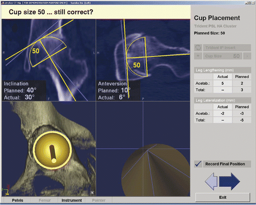
Figure 1. Screen display showing the inclination and anteversion of the cup, the anteversion and varus/valgus angle of the stem, and the leg length compared with the contralateral side.

Figure 2. Evaluation using postoperative CT. Each angle is displayed on the screen.

Figure 2. Evaluation using postoperative CT. Each angle is displayed on the screen.

Figure 3. Mean deviation of cup anteversion, inclination and stem anteversion using navigation and manual methods. There is only a significant difference for cup anteversion.
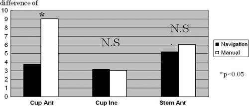
Figure 3. Mean deviation of cup anteversion, inclination and stem anteversion using navigation and manual methods. There is only a significant difference for cup anteversion.

Figure 4. Mean deviation of leg length compared with the contralateral side using navigation. 52 of the 56 cases are within 5 mm, and 41 of the cases are within 2 mm.
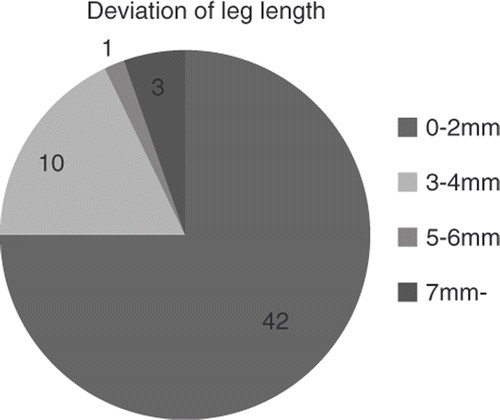
Figure 4. Mean deviation of leg length compared with the contralateral side using navigation. 52 of the 56 cases are within 5 mm, and 41 of the cases are within 2 mm.
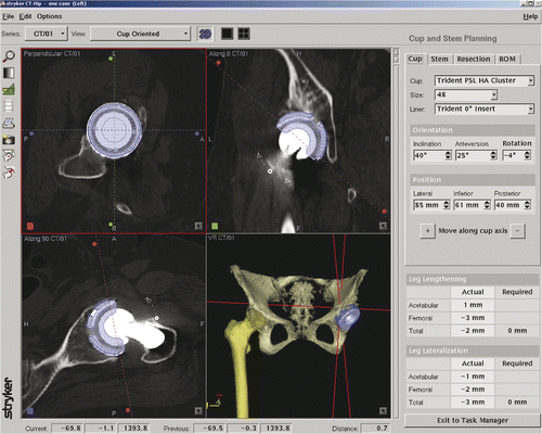
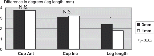
Figure 5. Mean deviation of cup inclination, cup anteversion and leg length using 3-mm and 1-mm CT slices. The deviation of the two cup parameters showed no significant difference.

Figure 5. Mean deviation of cup inclination, cup anteversion and leg length using 3-mm and 1-mm CT slices. The deviation of the two cup parameters showed no significant difference.

Figure 6. Although the varus angle of this stem is accurate to 3°, it appears to be ill-shapen on the X-ray. This demonstrates that accurate numerical values for the varus/valgus angle do not guarantee good results.

Figure 6. Although the varus angle of this stem is accurate to 3°, it appears to be ill-shapen on the X-ray. This demonstrates that accurate numerical values for the varus/valgus angle do not guarantee good results.

Reprints and Corporate Permissions

Please note: Selecting permissions does not provide access to the full text of the article, please see our help page How do I view content?

To request a reprint or corporate permissions for this article, please click on the relevant link below:

Academic Permissions

Please note: Selecting permissions does not provide access to the full text of the article, please see our help page How do I view content?

Obtain permissions instantly via Rightslink by clicking on the button below:

If you are unable to obtain permissions via Rightslink, please complete and submit this Permissions form. For more information, please visit our Permissions help page.