Figures & data
Figure 1. Dorsal view of the femoral intercondylar notch with the areas three-dimensionally digitized by Fastrak: (I) the outlines of the AM (white stars) and PL (black stars) ligament insertion sites; (II) the surface of the lateral notch (including the insertion areas); and (III) the cartilage line of the posterior joint cartilage edge at the medial wall of the lateral condyle (black dots).
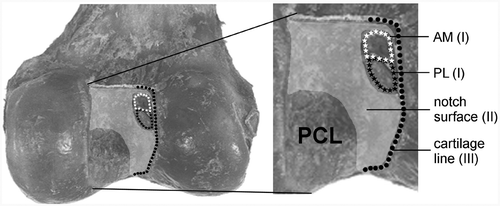
Figure 2. The notch surface in the dorsal view (A), lateral view (B) and notch view (C), which can be approximated by a cylindrical shape. The radius, orientation and position of the cylinder are determined by the roof points (A). The cylinder position in the proximal-distal direction (B) and the rotation along the longitudinal axis (C) were determined by the origin (0, 0) of the cylindrical coordinate systems (also see ).
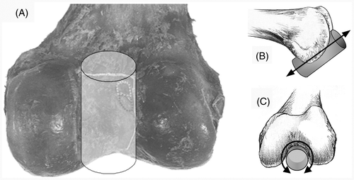
Figure 3. (A) Ventrolateral view of the fitted cylinder with the projected data of the cartilage border and the AM and PL insertion centers. A parabolic curve was fitted through the points of the cartilage border. The top of the parabola was defined as the origin (0, 0) at the cylindrical surface. (B) From the mean center points, the distance in the axial direction (Z) in mm and the angle φ were defined. The direct distance (2D vector D) between the centers and the origin was calculated with Pythagoras’ theorem, with XD being the result of sin 0.5φ*R*2.
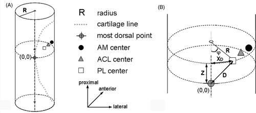
Figure 4. (A) Digitizing the notch surface points with the computer-tracked Stille hook. The femur is tracked through the dynamic reference base (DRB). (B) The digitized notch surface is visualized in three dimensions with a Levenberg-Marquardt algorithm for better orientation. The CAS system is calculating the cylinder-algorithm to fit the notch surface. Left: notch view; right: lateral-medial view.
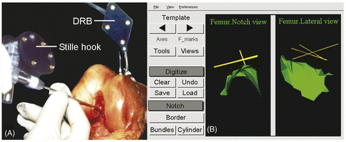
Figure 5. (A) Digitizing the cartilage line points at the medial notch wall of the lateral femoral condyle with the tip of the Stille hook. (B) The CAS system positions the scaled template with the anatomic red AM, blue PL and white ACL centers. Left: notch view; right: lateral-medial view.
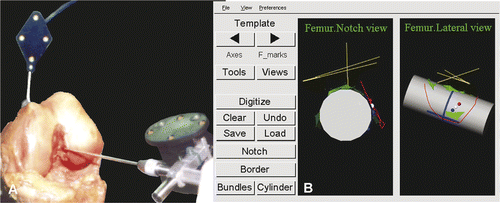
Figure 6. (A) Marking of the drill-hole location in the notch with a physical indent of the computer-tracked awl. (B) The red anatomic AM center is the target point for the mark by the transparent pedicle awl, as visualized on the CAS system monitor. Left: notch view; right: lateral-medial view.
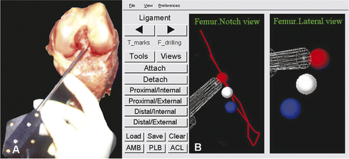
Figure 7. Three-dimensional coordinate system as defined in the SurgiGATE CAS system for the accuracy and precision measurements of the femoral template. The axes of the femur were defined by digitizing points with the Stille hook. The long Z-axis (white) between the distal point in the middle of the condyles and the middle of the shaft 10 cm proximal above the knee, where the femur was cut, is comparable to the shallow-deep direction in the notch. (This is in contrast with the regular situation, where the trochantor major femoralis is digitized.) The medial-lateral Y-axis (gray line) between the two femoral epicondyles, perpendicular to the notch wall, will not be taken into account, since the tunnel is placed at the bony surface. The X-axis (black line) follows from the other two axes and runs from ventral to dorsal, comparable to the high-low direction in the notch.
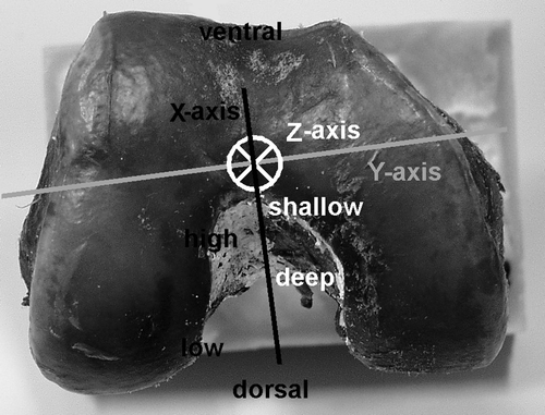
Figure 8. Diagram of the points on the cylinder surface (left) and a lateral view of a femur (with the lateral condyle dissected) (right) showing the alignment of the 3D cylinder template including the projections on the surface of the origin of the coordinate system (the apex of the cartilage border) and the mean position of the centers of the ACL, AM and PL. Z is the distance in the shallow-deep direction; X is the distance in the low-high direction. The error bars represent 1.96*SD, containing 95% of the individual centers (95% CI).
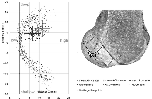
Table I. Mean anatomical positions and variations (SD) of the insertion centers in the template, relative to the apex of the cartilage border.
Figure 9. The positions of the template centers relative to the anatomic centers (bull's eye) for AM (A) and PL (B). The circles indicate the distance in mm. The total error is composed of the accuracy, represented by the distances in the high-low and shallow-deep directions between the bull's eye and the mean template centers, and the precision, represented by 1.96*SD (black lines).
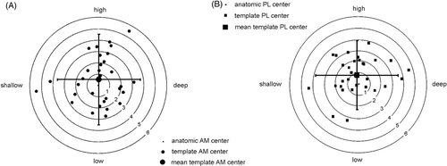
Table II. Accuracy and precision of the tunnels placed by the template using the CAS system, relative to the anatomical centers.