Figures & data
Figure 1. Operative set-up showing the dynamic reference base fixed on the distal femur and image guided freehand navigation of the guide wire into the femoral neck.
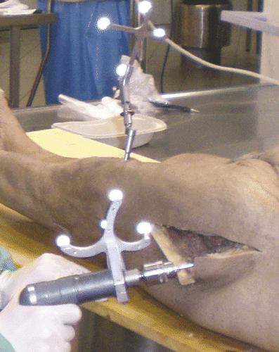
Figure 2. Field of view (FOV) registration points (in green) defined by the surgeon to determine the reference frame for his eye-hand coordination. The blue point represents the tip of the guide pin.

Figure 3. Image acquisition with zero-dose C-arm navigation. (a) Preview of frontal view (with acquired image in background). (b) Preview of lateral view (with acquired image in background).
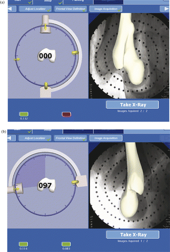
Figure 4. Definition of the safe zone: planning the implant position with the displayed safety cylinder (blue cylinder) and safety sphere (red sphere) in the frontal view (a) and lateral image (b).
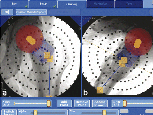
Figure 5. Planning of the screw position in the frontal (left) and lateral (right) image in the femoral neck without exceeding the femoral head.
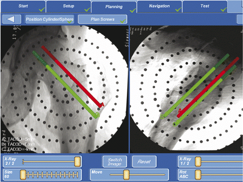
Figure 6. On leaving the safe zone the surgeon is warned by the appearance of the “Perforation” caption.
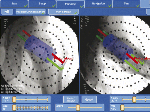
Figure 7. (A) Visualization of the image guided freehand navigation of the guide wire on the planned trajectory. (B) Display of two crosshair-like vectors for orientation of the navigation tool. The green circle (a) represents the position of the guide wire tip, and the red diamond (b) represents the position of a point on the guide wire axis. (c) is the crosshair-like representation of the online distance and direction from the guide wire tip to the axis of the planned trajectory (the diamond and circle in the center mean that the tool is aligned). (d) is a representation of the depth of the guide wire in relation to the lateral cortical bone.
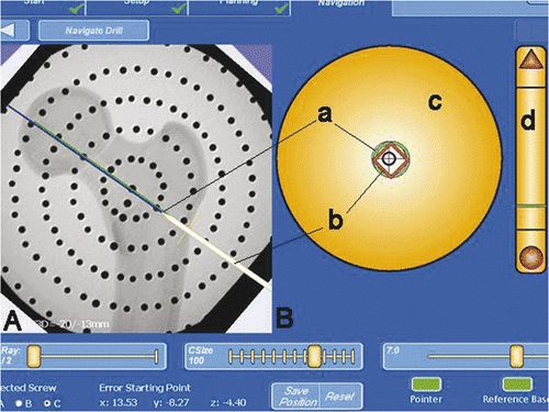
Figure 8. Screenshot of the final implant position in the frontal (a) and lateral (b) views.
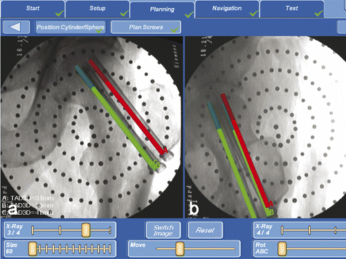
Table I. Comparison of the conventional fluoroscopic technique (Group 1) with the computer-assisted technique (Group 2). The values are given as the mean and standard deviation. Statistical significance was defined for *p < 0.05; **p < 0.001.
Table II. Comparison of 2D fluoroscopy-based navigation systems for navigating cannulated femoral neck screw placement. (CAS: Computer Assisted Surgery; COT: conventional operative technique. Statistical significance: *p < 0.05; **p < 0.001).