Figures & data
Figure 1. Methodology for registering pre-operative CT to intra-operative ultrasound.
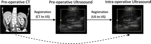
Figure 2. Conventional tracked ultrasound approach for US-to-CT registration.
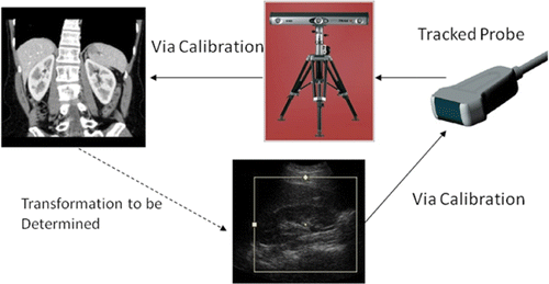
Figure 3. Linear-array ultrasound beam pattern.
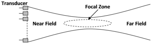
Figure 4. Prototype stand-off pad with five fiducial markers.
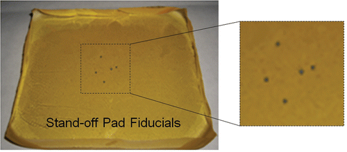
Figure 5. Schematic of testing phantom containing phantom target fiducials (fiducial stand-off pad is secured to top).
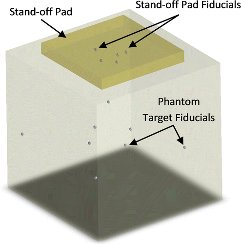
Figure 6. Sample ultrasound image showing one phantom fiducial and two stand-off pad fiducials.
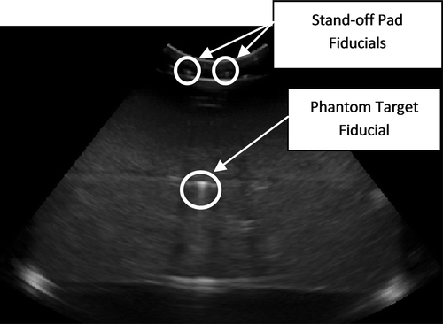
Figure 7. Sample CT image showing one phantom fiducial and one stand-off pad fiducial. The curved appearance of the stand-off pad is due to the coupling gel between the pad and phantom.
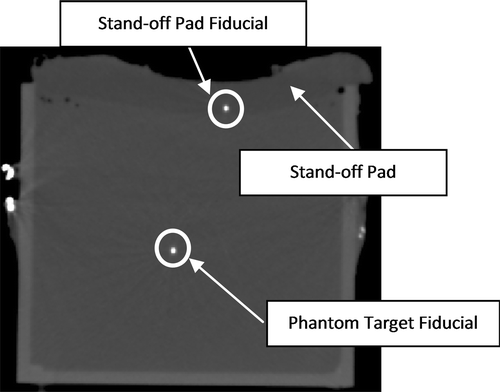
Table I. Ultrasound scanning parameter sets. Volumes contained 76 frames (55° sweep).
Figure 8. Demonstration of the lever arm effect. White fiducials represent the true locations, and black fiducials represent locations with localization error. A small misalignment of the stand-off pad fiducials can result in large target registration errors.
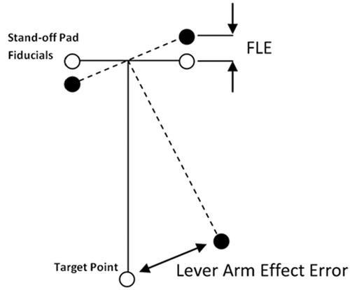
Figure 9. The CIRS quality assurance phantom with no stand-off pad (left), with a 12-mm thick stand-off pad (center), and with a 20-mm thick stand-off pad (right). Ultrasound acquisition settings were as follows: probe frequency 5 MHz; focal depth 7.6 cm; depth 15 cm; gain 48%. The arrows show the point feature target number 1, with numbers 2 to 9 below it in sequence, as in . The circles show the occlusion feature number 1, with numbers 2 and 3 diagonally below it in sequence, as indicated in .
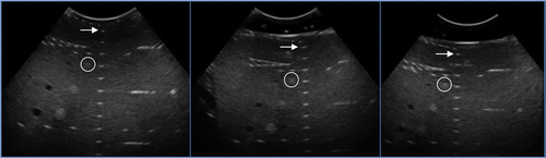
Figure 10. Sample image of line features used to determine the degradation of resolution in the elevational direction. The arrow shows line feature target number 1, with numbers 2 to 9 below it in sequence (as in ).
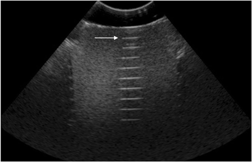
Table II. Effect of stand-off pad thickness on image quality. Ultrasound acquisition settings were as follows: frequency 5 MHz; focal depth 7.6 cm; depth 15 cm; gain 48%. There were 76 frames/volume (55° sweep). Point feature numbering is indicated in .
Table III. Effect of stand-off pad thickness on elevational resolution. Ultrasound acquisition settings were as follows: frequency 5 MHz; focal depth 7.6 cm; depth 15 cm; gain 48%. There were 76 frames/volume (55° sweep). Point feature numbering is indicated in .
Table IV. Effect of stand-off pad thickness on image contrast. Ultrasound acquisition settings were as follows: frequency 5 MHz; focal depth 7.6 cm; depth 15 cm; gain 48%. Occlusion feature numbering is indicated in .
Figure 11. FLE vs. various ultrasound parameters (see ). In each panel, the left graph shows errors in measurements of distances between stand-off pad fiducials (N = 6) indicating lateral and elevational errors, and the right graph shows errors in measurement of distances between the stand-off pad and phantom target fiducials (N = 32) indicating axial errors. (a) FLE vs. probe frequency; (b) FLE vs. focal depth; (c) FLE vs. gain; and (d) FLE vs. depth.
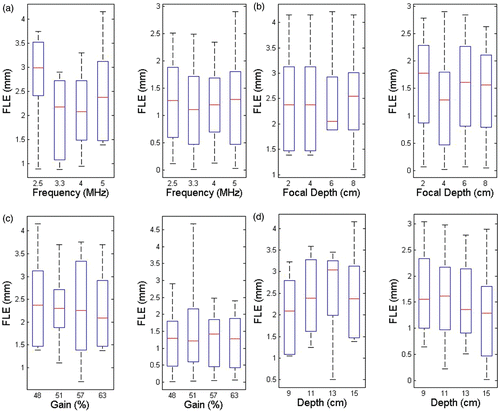
Figure 12. RMS FRE vs. various ultrasound parameters. (a) FRE vs. probe frequency (focal depth 4 cm; gain 48%; depth 15 cm). (b) FRE vs. focal depth (frequency 5 MHz; gain 48%; depth 15 cm). (c) FRE vs. gain (frequency 5 MHz; focal depth 4 cm; depth 15 cm). (d) FRE vs. depth (frequency 5 MHz; focal depth 4 cm; gain 48%).
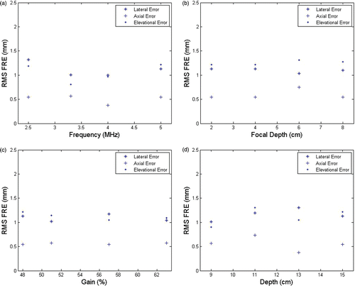
Figure 13. Absolute FRE in the lateral, axial and elevational directions for all datasets (see ).
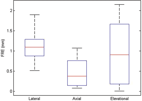
Figure 14. RMS TRE vs. various ultrasound parameters. (a) TRE vs. probe frequency (focal depth 4 cm; gain 48%; depth 15 cm). (b) TRE vs. focal depth (probe frequency 5 MHz; gain 48%; depth 15 cm). (c) TRE vs. gain (probe frequency 5 MHz; focal depth 4 cm; depth 15 cm). (d) TRE vs. depth (probe frequency 5 MHz; focal depth 4 cm; gain 48%).
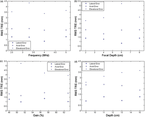
Figure 15. Absolute TRE in the lateral, axial and elevational directions for all data sets (see ).
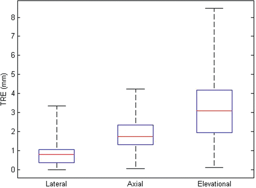
Figure 16. Sample CT (left) and ultrasound (right) images of patient's kidney with stand-off pad attached. Ultrasound acquisition settings were as follows: frequency 5 MHz; focal depth 7.6 cm; gain 42%; depth 13 cm.
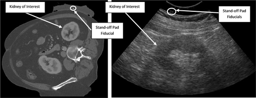
Figure 17. Overlays of clinical data for three patients after registration using the fiducial stand-off pad. CT data is shown in blue and ultrasound data in red. (a) Patient 1; (b) Patient 2; (c) Patient 3.
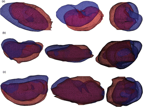
Table V. RMS FRE for clinical data from three patients.
Table VI. TRE for clinical data from three patients.
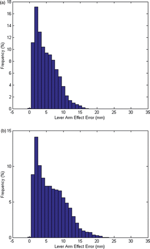
Figure 18. Histograms of lever arm errors. Bin size = 1 mm; N = 32768. (a) For a target point 60 mm from the fiducials to simulate the phantom study. (b) For a target point 80 mm below the fiducials to simulate the clinical setting.