736
Views
12
CrossRef citations to date
0
Altmetric
Brief Report

A method for three-dimensional evaluation and computer aided treatment of femoroacetabular impingement

, &
Pages 143-148 | Received 30 Nov 2010, Accepted 10 Jan 2011, Published online: 28 Mar 2011

Figures & data

Figure 1. (A) Craniocaudal view of femur with cam-type lesion (dark blue area) as detected by morphological analysis, and (B) suggested surgical correction to be obtained (red area).
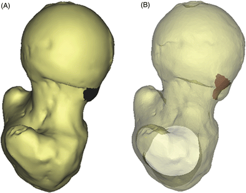
Figure 1. (A) Craniocaudal view of femur with cam-type lesion (dark blue area) as detected by morphological analysis, and (B) suggested surgical correction to be obtained (red area).

Figure 2. (A) Description of femoral and acetabular contact points in relation to the location and size of the cam lesion, and (B) identification of the intra-articular collision area and contre-coup area for evaluation of cartilage at risk for shear damage during specific motions (red areas). This particular example represents a simulation for flexion in a clinical case of FAI.
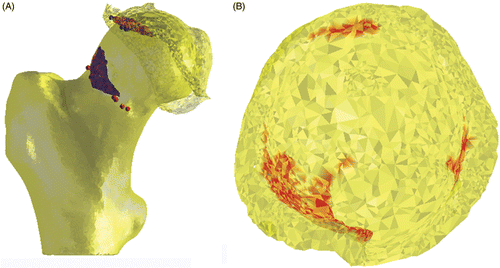
Figure 2. (A) Description of femoral and acetabular contact points in relation to the location and size of the cam lesion, and (B) identification of the intra-articular collision area and contre-coup area for evaluation of cartilage at risk for shear damage during specific motions (red areas). This particular example represents a simulation for flexion in a clinical case of FAI.

Figure 3. Structural identification of a pincer-type lesion by means of penetration evaluation of the reshaped femur in the acetabular rim during combined motions.
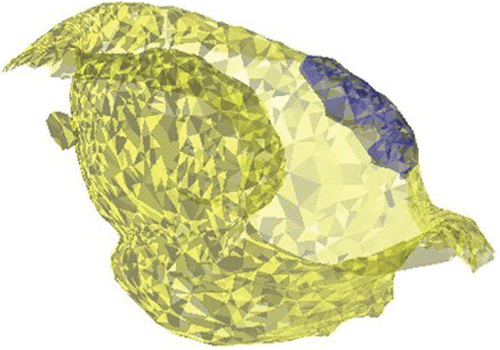
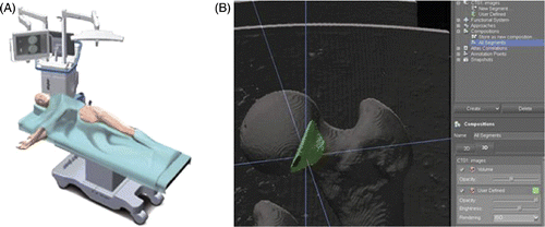
Figure 3. Structural identification of a pincer-type lesion by means of penetration evaluation of the reshaped femur in the acetabular rim during combined motions.
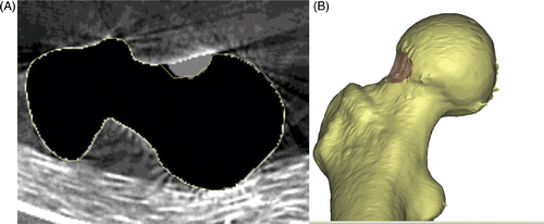
Figure 4. Navigated surgical resection of the cam lesion based on structural analysis of the femur. (A) Patient set-up and (B) graphical user interface, including 3D planning tools that provide information on the size of the lesion and the location of the surgical tool in relation to it.

Figure 4. Navigated surgical resection of the cam lesion based on structural analysis of the femur. (A) Patient set-up and (B) graphical user interface, including 3D planning tools that provide information on the size of the lesion and the location of the surgical tool in relation to it.

Figure 5. In vitro evaluation of computer-assisted surgical accuracy. (A) Axial CT reconstruction of the planned (grey area) versus actual (yellow outline) resection of a cam-type lesion in a sawbone model; and (B) 3D reconstruction of the postoperative result.

Figure 5. In vitro evaluation of computer-assisted surgical accuracy. (A) Axial CT reconstruction of the planned (grey area) versus actual (yellow outline) resection of a cam-type lesion in a sawbone model; and (B) 3D reconstruction of the postoperative result.

Reprints and Corporate Permissions

Please note: Selecting permissions does not provide access to the full text of the article, please see our help page How do I view content?

To request a reprint or corporate permissions for this article, please click on the relevant link below:

Academic Permissions

Please note: Selecting permissions does not provide access to the full text of the article, please see our help page How do I view content?

Obtain permissions instantly via Rightslink by clicking on the button below:

If you are unable to obtain permissions via Rightslink, please complete and submit this Permissions form. For more information, please visit our Permissions help page.