Figures & data
Figure 1. Three-dimensional visualization of the CT volume data and illustration of the original position of PClaser in the image space. The green lines in the left and right panels are the original positions of PClaser1 and PClaser2, respectively. In each case, the portion of the green line falling within the red rectangle is the area where anisotropic errors were added.
Figure 2. The position of the TRE sampling points. The red, green and blue lines are the X′, Y′ and Z′ sampling line segments, respectively. They intersect at a point at the center of the head, and are parallel to the X, Y and Z coordinate axes of the volume data.
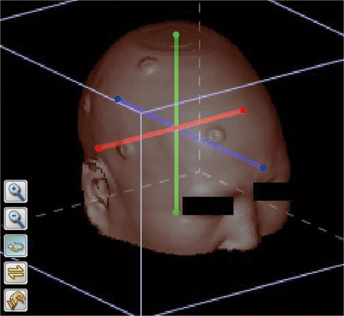
Table I. Generation method and number of point clouds in the patient space.
Figure 3. Spatial distribution of TRE in surface matching. The left set of images represent a case of registration with scanning area PClaser1. The right set of images represent a case of registration with scanning area PClaser2. In both cases, the upper left, upper right and lower left panels correspond to the coronal, sagittal and axial sections that pass through the center point, which is indicated by a small red cross. The lower right panel in each set is the 3D visualization of the head segmented out from the background by thresholding. On the section images, the TRE at the points where the CT value is larger than the threshold is mapped into a gray value, with dark gray indicating a smaller TRE and light gray indicating a larger TRE. The TRE in the area within the innermost iso-valued surface is 0.5 voxels in the left image and 1.6 voxels in the right image, and the TRE increases by 0.1 voxels with each iso-valued surface moving outward.
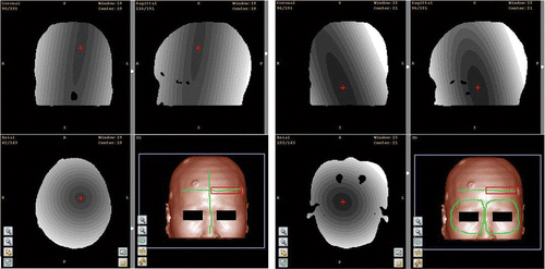
Figure 4. Statistical distribution of TRE on sampling axes with different scanning areas and error types. Small dots and triangles indicate the mean values of the TRE for PClaser1 and PClaser2, respectively, and the horizontal bars indicate the standard deviation. IE, SAE and LAE are the three types of errors: isotropic random error, small anisotropic random error, and large anisotropic random error. (a), (c) and (e) show the TRE distributions on the X′, Y′ and Z′ sampling axes when PClaser1 was used. (b), (d) and (f) show the TRE distributions on the X′, Y′ and Z′ sampling axes when PClaser2 was used.
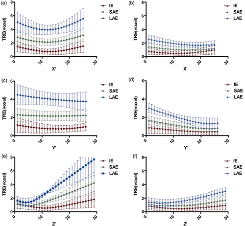
Figure 5. Statistical distribution of SRE for registrations with different scanning areas and error types. Small dots indicate the mean value of the SRE for registrations with PClaser1, and small triangles indicate the mean value of SRE for registrations with PClaser2. The horizontal bars indicate the standard deviation. IE, SAE and LAE are the three types of error: isotropic random error, small anisotropic random error, and large anisotropic random error.
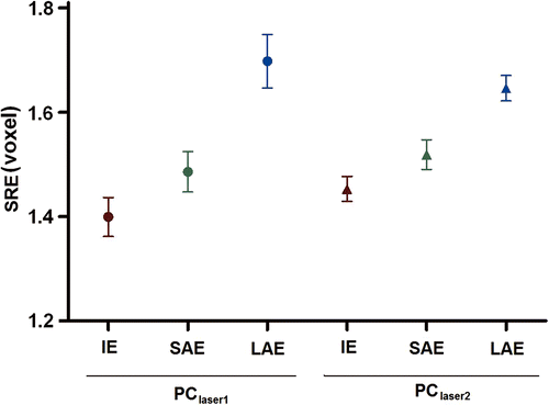
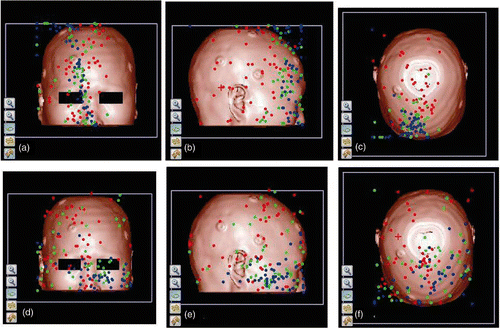
Figure 6. Spatial distribution of the center points for registrations with different scanning areas and error types. Fifty registrations were performed for each combination of scanning area and error type. The red, green and blue dots indicate the position of the center points for IE, SAE and LAE, respectively. (a), (b) and (c) are the distributions for the scanning area PClaser1, viewed from the front, right and top, respectively. (d), (e) and (f) are the distributions for the scanning area PClaser2, viewed from front, right and top, respectively.