Figures & data
Figure 1. 2D projection of a 3D model.
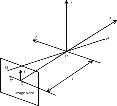
Figure 2. (a) Volume-rendered phantom brain CT image and (b) surface-rendered brain model.
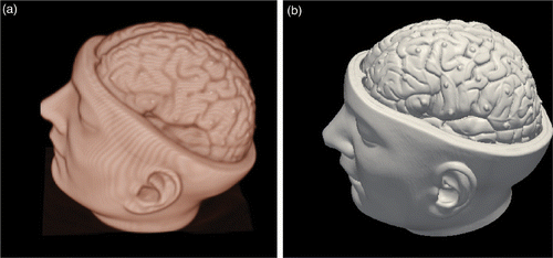
Figure 3. Phantom experiment. (a) Landmarks on the CT image. (b) Landmarks on the photograph.
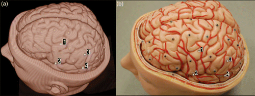
Figure 4. Result of phantom experiment. (a) Photographic overlay on the CT image. (b) Magnification of the region of interest in (a).
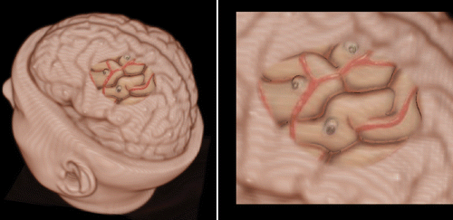
Table I. Image fusion results in the phantom experiment.
Figure 5. Clinical evaluation: paper tags on the cortical surface.
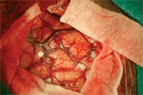
Figure 6. Clinical evaluation: landmarks used to align the two images initially. (a) Landmarks defined on the volume-rendered image. (b) Landmarks defined in the 2D photographic image.
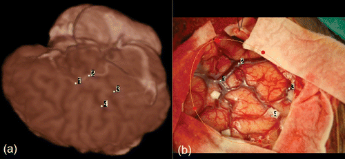
Figure 7. Clinical evaluation. (a) Fusion of the photographic image onto the MR brain model and reconstructed landmarks in the clinical study. (b) Magnification of the region of interest in (a).
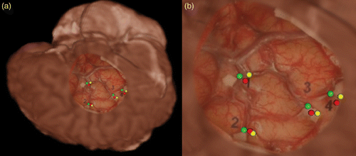
Table II. Image fusion results in the clinical experiment.
Table III. Brain shift measurements in the clinical experiment.
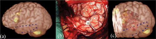
Figure 8. Clinical implementation. (a) Volume rendering of the anatomical MR volume and functional activation map. (b) Intraoperative photograph of left temporal lobe. (c) Overlay of the photograph on the volume rendering of the anatomical MR volume and functional activation map.