Figures & data
Figure 1. Navigation system overview. The transformations camTndr and pdrTcam are measured by the position sensor, whereas imgRpdr is the patient-to-image registration.
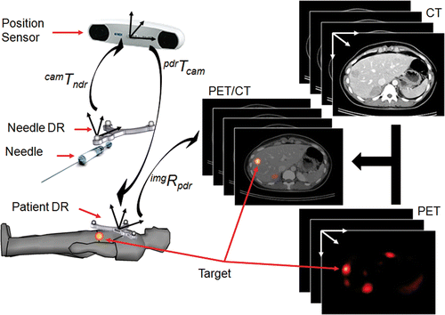
Figure 2. (a) 3D viewer showing an overview of the planned situation and the orientation of the trajectory viewer. (b) Trajectory viewer showing the planned trajectory and possible obstacles. (c) Navigation platform beside the PET/CT scanner table. (d) Needle and calibration unit in calibration position.
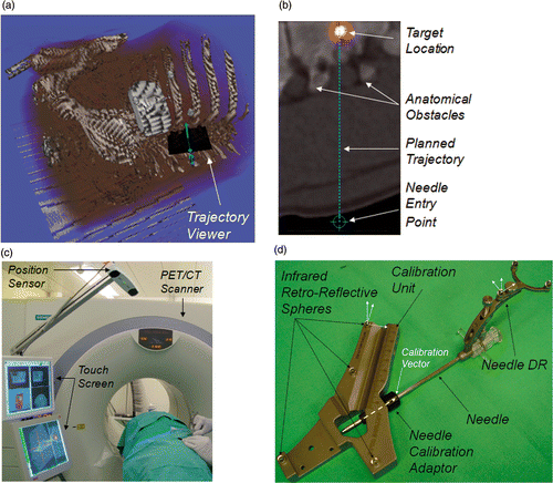
Figure 3. (a) Schematic of the targeting viewer. The virtual camera is aligned with the planned trajectory and looks towards the target. The needle tip and the needle shaft are projected back to the crosshair plane, and are respectively displayed as a cross and a circle that is complementary to the cross. Both are connected through a line. The viewer background displays the fused PET/CT slice orthogonal to the planned trajectory at the depth of the needle tip d, or alternatively 10 mm beyond the tip, d + 10. (b) Schematic of the needle plane viewer. The virtual camera looks towards the needle tip, and has its y axis parallel to the needle, and its x axis parallel to the cross-product between the needle and the z axis of the needle DR. The viewer background displays the PET/CT slice surrounding the needle axis. To aid the physician in predicting the final tip position if the needle continues in the current direction, a projection of the needle along the remaining distance d is displayed.
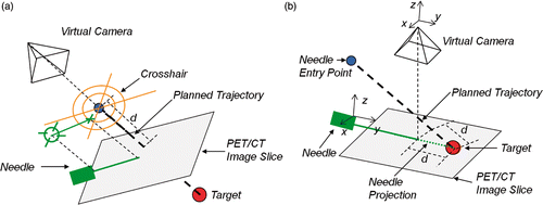
Figure 4. (a) The targeting viewer with the needle aligned with the planned trajectory and moving towards the target. The distance to the target is displayed in the left bar, and is also represented by the distance arrows approaching the center of the cross-hair target, which removes the necessity to deviate one's attention from the center of the viewer. (b) The needle plane viewer displaying the needle, its projection to the target, and the PET/CT slice along the needle axis.
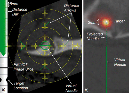
Figure 5. System workflow emphasizing the non-sterile and sterile phases and the particularities of bone and soft tissue interventions.
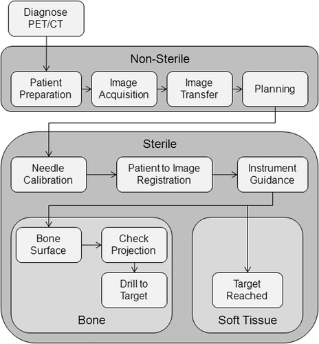
Figure 6. Illustration of the target projection and error components. (a) Different projection methods: patient DR registration transformation Rpdr, rigid structure registration transformation Rrig, and machine coordinate system transformation Rmcs. (b) Locations of the target projected with different methods: patient DR registration transformation RpdrPplt, rigid structure registration transformation RrigPplt, and machine coordinate system transformation RmcsPplt. Additionally, the picture on the right illustrates how the different error vectors correlate to one another.
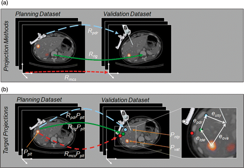
Figure 7. (a) The best result for the bone punctures: puncture 14 in S1 with a planned trajectory length of 81 mm. (b) The best result for the soft tissue punctures: puncture 2 in S3 with a planned trajectory length of 98 mm.
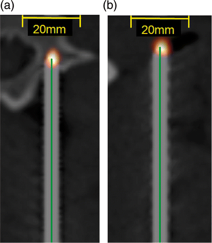
Table I. Average and standard deviation of the error sources (in mm).
Figure 8. (a) and (b) show the different error sources per puncture for bone and soft tissue punctures, respectively, ordered by the overall error. (c) and (d) show the correlation between the error sources and the overall error for bone and soft tissue punctures, respectively. The lines represent the linear fitting of each error source.
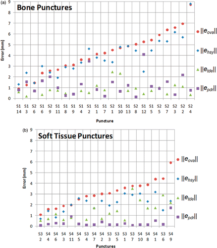
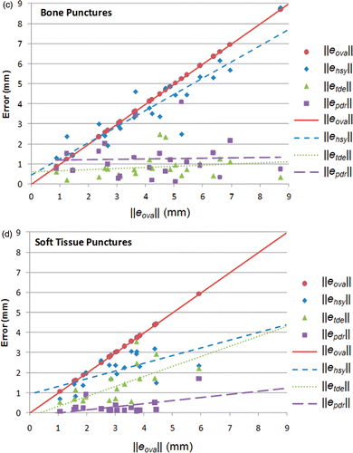
Figure 9. Results of punctures in S2 with high error in the drilling direction: (a) puncture 2; (b) puncture 3; (c) puncture 5; and (d) puncture 7.
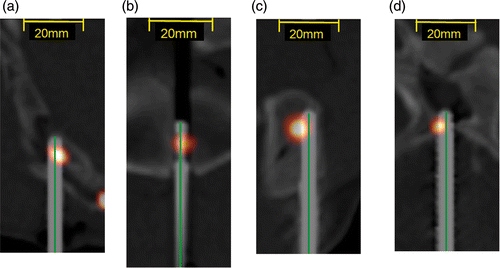
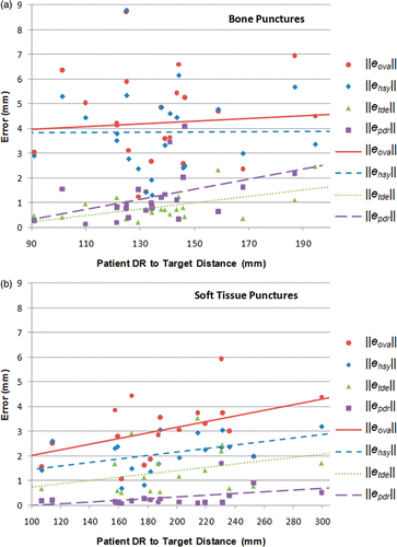
Figure 10. The different error sources per (a) patient DR to target distance for bone punctures; (b) patient DR to target distance for soft tissue punctures; (c) trajectory length of the bone punctures; and (d) trajectory length of the soft tissue punctures. The lines represent the linear fitting of each error source.