Figures & data
Figure 1. (a) The geometric phantom was a Styrofoam cube with 25 fiducial markers, each consisting of a metal ball 1.5 mm in diameter, fixed in a cross arrangement at intervals of 1 cm. (b) A 2-dimensional image from the C-arm.
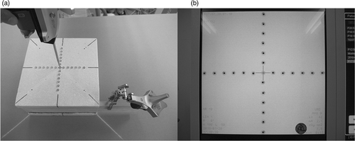
Figure 2. Experimental set-up of the geometric phantom study (a) and dry bone study (b). The C-arm was placed on the left side of the operation table and the navigation system was placed at the caudal end. In the geometric phantom study, the plane of the phantom with the markers affixed faced upward.
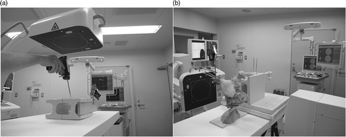
Figure 3. (a) Surgery around the hip joint was simulated with a dry human pelvis and femur placed on the operating table in the lateral decubitus position. (b) A 2-dimensional image from the C-arm.
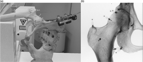
Figure 4. The TRE of markers located at 5 cm from the imaging center was higher than the TRE of markers located at 1 and 2 cm. TRE was <1 mm in 90% of the overall trials and <1.5 mm in 100%.
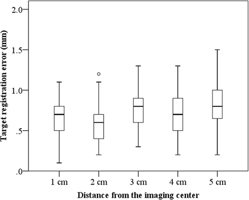
Figure 5. No significant differences in TRE were seen between the acetabulum and proximal femur.
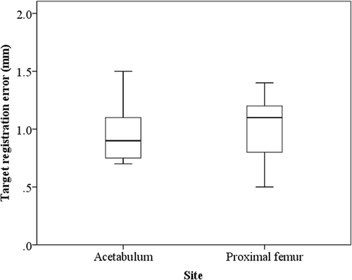
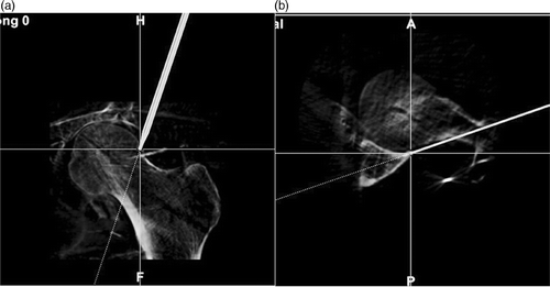
Figure 6. Visual assessment of navigation accuracy was performed by indicating several landmarks with the navigation probe. Mutiplanar images of the head-neck junction of the femur (a) and posterior acetabular edge (b) are shown.