Figures & data
Figure 1. The experimental set-up for mechanized pivot shift testing. The lower limb is mounted in full extension on the mechanized pivot shifter, abducted by at least 15° at the hip joint. The foot is affixed to the base plate and is externally rotated. The base plate can then be pushed by the examiner along two linear bearing surfaces, effectively cycling the knee from 0° to 90° of flexion. Using a load cell attached to the mechanized pivot shifter through a three-degrees-of-freedom arm, a valgus load of 4–5 kg is applied to the proximal third of the tibia to reproduce the pivot shift phenomenon. The computer navigation system tracks the tibiofemoral motion path using a camera that emits and then captures a signal reflected by the passive reflective markers attached to the femur and tibia. The arc of motion during the pivot shift test is compared to a flexion-extension path acquired during the registration process of the intact knee and is then visually represented on the screen.
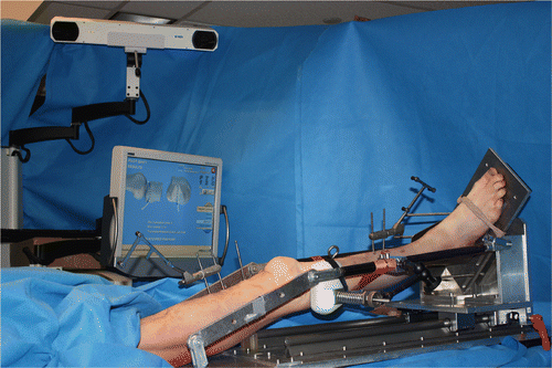
Figure 2. Screenshot of the Pivot Shift Visualizer interface. Once the bone morphing and kinematic data files are loaded, the program displays a 3D rendering of the tibia and the femur, along with a coordinate system. The user may then replay the tibiofemoral motion recorded by the navigation system during the pivot shift test.
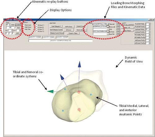
Figure 3. During kinematic replay, the tibiofemoral translation path is represented by yellow dots connected by green lines. This visual representation aids the examiner in tracking the motion of the tibia over the femur during each test. The bones may be rotated through 360° to allow visualization from any angle (A, B).
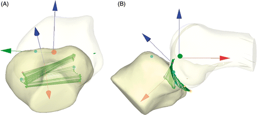
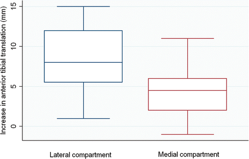
Figure 4. The increase in tibiofemoral translation after sectioning the ACL was significantly greater in the lateral compartment compared to the medial compartment (p < 0.05).