Figures & data
Figure 1. Case II. Axial (a), coronal (b) and sagittal (c) gadolinium-enhanced T1 MR sequences show an atrophic but clearly visible right optic nerve encased by a globular contrast-enhancing homogeneous lesion. The axial cut shows well the perineural growth pattern typical of ONMS, while the entire intraorbital course of the nerve can be appreciated. The same lesion appears on CT imaging [illustrated here in the context of the treatment plan (d)] as a large contrast-enhancing lesion encasing the right optic nerve.
![Figure 1. Case II. Axial (a), coronal (b) and sagittal (c) gadolinium-enhanced T1 MR sequences show an atrophic but clearly visible right optic nerve encased by a globular contrast-enhancing homogeneous lesion. The axial cut shows well the perineural growth pattern typical of ONMS, while the entire intraorbital course of the nerve can be appreciated. The same lesion appears on CT imaging [illustrated here in the context of the treatment plan (d)] as a large contrast-enhancing lesion encasing the right optic nerve.](/cms/asset/abce22b8-235c-4f20-a657-0641b7101d17/icsu_a_622615_f0001_b.gif)
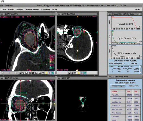
Figure 2. Treatment planning for patient I. CT imaging shows a contrast-enhancing globular lesion growing eccentrically to the left optic nerve, which is compressed and displaced. Treatment planning details such as dose delivered and DVHs are illustrated.
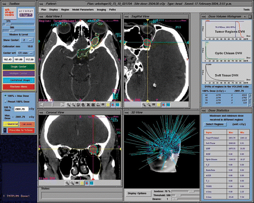
Figure 3. CyberKnife radiosurgery treatment planning for a right ONSM. This patient was a 28-year-old female presenting with progressive deterioration of visual fields and acuity in the right eye over the previous 6 months. (a) Optic nerve imaging was performed using 3-T MR (Siemens Trio). CT-MR fusion was performed on the CyberKnife treatment planning station. The nerve was identified as a loop displaced downward by the tumor and was drawn as a critical structure. This enhanced the degree of nerve sparing from irradiation. (b) A total of 109 beams were delivered non-isocentrically to the lesion. Treatment volume was 0.764 cc. A total dose of 20 Gy prescribed to the 70% isodose was delivered in 4 stages (with 5 Gy per stage) separated by an interval of 24 hours. Maximum dose was 28.57 Gy. Tumor and optic nerve dose volume histograms (DVHs) show a substantial sparing of the optic nerve.
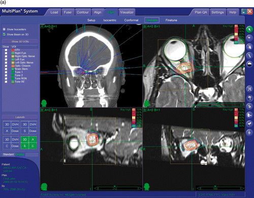
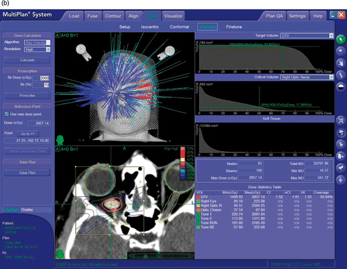
Figure 4. Visual fields before and after treatment for patient I. The upper row corresponds to the left eye, the lower row to the right eye. On each row, the first box is the visual field pre-treatment, the second box is the field after 6 months, and the third box is the field after 1 year. This patient maintained stable visual fields thereafter, with follow-up now approaching 8 years.
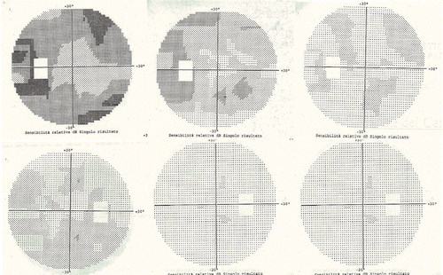
Table I. Patient and lesion characteristics, treatment parameters, outcome and follow-up duration.