Figures & data
Figure 1. Leg model with rigid tracker mountings.
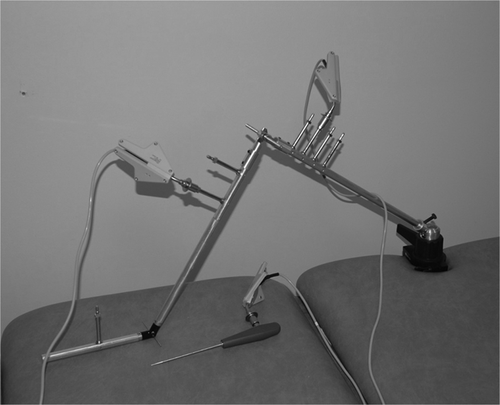
Figure 2. External tracker mountings with curved base plates and adjustable elasticated straps. (a) Mounting of trackers on thigh and calf. (b) Foot tracker mounting.
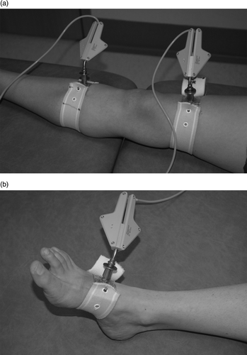
Table I. The mean and standard deviation (SD) of each set of tests was used to compare the difference in repeatability of the rigid leg model and the non-invasive tracker mounting on the volunteers.
Table II. Mean difference and 95% limits of agreement of repeat supine alignment measurements in extension with leg stationary and before and after both standing and collateral stress maneuvers (all measurements in degrees)
Table III. Inter-registration agreement of supine and standing coronal and sagittal MFT angles, and relative change following varus-valgus stress (measurements in degrees).
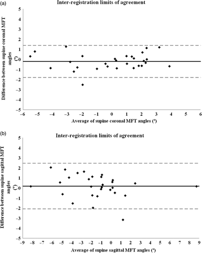
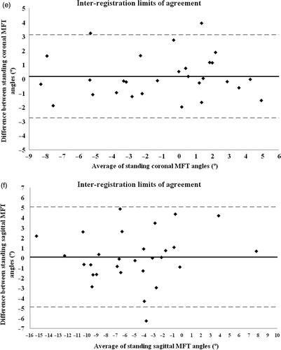
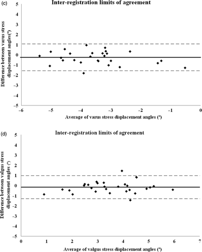
Figure 3. Bland-Altman plots showing mean difference (solid black line) and 95% limits of agreement (dotted grey lines) of MFT angular measurements for two trials: (a) supine coronal; (b) supine sagittal; (c) with varus stress; (d) with valgus stress; (e) standing coronal; and (f) standing sagittal.