763
Views
3
CrossRef citations to date
0
Altmetric
Research Article

Three-dimensional multimodal image non-rigid registration and fusion in a High Intensity Focused Ultrasound system

, Ph.D., , &
Pages 1-12 | Received 25 Apr 2011, Accepted 04 Oct 2011, Published online: 08 Dec 2011

Figures & data

Figure 1. High Intensity Focused Ultrasound (HIFU) therapy system.

Figure 1. High Intensity Focused Ultrasound (HIFU) therapy system.

Figure 2. Flowchart of the image-guided localization system in our HIFU.

Figure 2. Flowchart of the image-guided localization system in our HIFU.

Figure 3. (a) Therapy probe in HIFU system. (b) B-mode ultrasound imaging probe in HIFU system.

Figure 3. (a) Therapy probe in HIFU system. (b) B-mode ultrasound imaging probe in HIFU system.

Figure 4. (a) The kidney region delineated on an MR image. (b) 3D visualization of therapy targeting fractions. The red, green and blue regions represent three different targeting fractions.
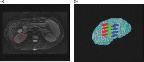
Figure 4. (a) The kidney region delineated on an MR image. (b) 3D visualization of therapy targeting fractions. The red, green and blue regions represent three different targeting fractions.

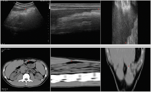
Figure 5. The three-view user interface including ultrasound and CT volume data.

Figure 5. The three-view user interface including ultrasound and CT volume data.

Table I.  Affine registration results and parameters of true deformation.

Figure 6. Registration in the simulated data experiment. (a) Reference image. (b) Floating image. (c) Registration result for the ROI using PV. (d) Difference image between (c) and the ROI of (a). (e) Registration result for the ROI using HPV. (f) Difference image between (e) and the ROI of (a).
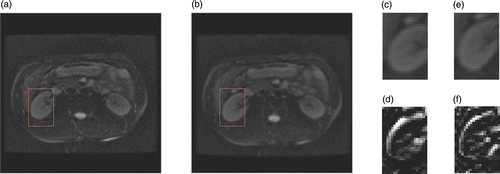
Figure 6. Registration in the simulated data experiment. (a) Reference image. (b) Floating image. (c) Registration result for the ROI using PV. (d) Difference image between (c) and the ROI of (a). (e) Registration result for the ROI using HPV. (f) Difference image between (e) and the ROI of (a).

Table II.  Comparison of registration results with known warping.

Figure 7. The distribution of the metric values with iterations after PV and HPV interpolation algorithms. (a) Using PV interpolation. (b) Using HPV interpolation.
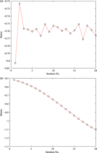
Figure 7. The distribution of the metric values with iterations after PV and HPV interpolation algorithms. (a) Using PV interpolation. (b) Using HPV interpolation.

Figure 8. Registration between CT and ultrasound images of the liver. (a) Reference image in axial (top), sagittal (middle) and coronal (bottom) planes. (b) Floating image in axial, sagittal and coronal planes. (c) Fused image after affine registration in axial, sagittal and coronal planes. (d) Deformed floating image after local non-rigid registration in axial, sagittal and coronal planes.
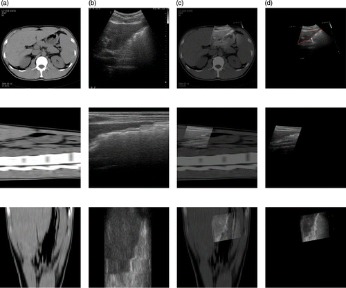
Figure 8. Registration between CT and ultrasound images of the liver. (a) Reference image in axial (top), sagittal (middle) and coronal (bottom) planes. (b) Floating image in axial, sagittal and coronal planes. (c) Fused image after affine registration in axial, sagittal and coronal planes. (d) Deformed floating image after local non-rigid registration in axial, sagittal and coronal planes.
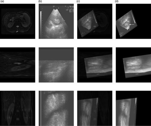
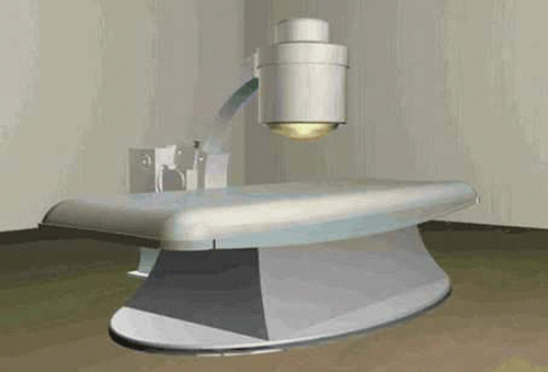
Figure 9. Registration between MR and ultrasound images of the kidney. (a) Reference image in axial (top), sagittal (middle) and coronal (bottom) planes. (b) Floating image in axial, sagittal and coronal planes. (c) Fused image after affine registration in axial, sagittal and coronal planes. (d) Deformed floating image after local non-rigid registration in axial, sagittal and coronal planes.
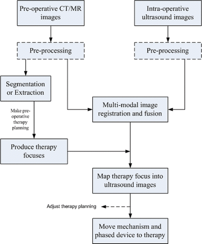
Figure 9. Registration between MR and ultrasound images of the kidney. (a) Reference image in axial (top), sagittal (middle) and coronal (bottom) planes. (b) Floating image in axial, sagittal and coronal planes. (c) Fused image after affine registration in axial, sagittal and coronal planes. (d) Deformed floating image after local non-rigid registration in axial, sagittal and coronal planes.
Reprints and Corporate Permissions

Please note: Selecting permissions does not provide access to the full text of the article, please see our help page How do I view content?

To request a reprint or corporate permissions for this article, please click on the relevant link below:

Academic Permissions

Please note: Selecting permissions does not provide access to the full text of the article, please see our help page How do I view content?

Obtain permissions instantly via Rightslink by clicking on the button below:

If you are unable to obtain permissions via Rightslink, please complete and submit this Permissions form. For more information, please visit our Permissions help page.