Figures & data
Figure 1. Fiducial configuration. (A) Four fiducial markers were attached to the humerus, two medially and two laterally, both proximally and distally around the joint articulation. A fifth fiducial was positioned on the articulation (region of interest) to measure TRE. (B) Four fiducial markers were attached to the ulna, one on the olecranon, one on the coronoid, and two distal markers on the medial and lateral side. A fifth fiducial was also positioned on the greater sigmoid notch (area of interest) to measure TRE.
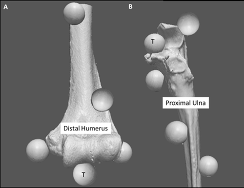
Figure 2. Ulnar subchondral zones. Four zones were created by dividing the ulnar subchondral bone medially and laterally down the ridge of the greater sigmoid notch (extending from the olecranon to the coronoid process). A second plane was created along the transverse ridge dividing the ulna into superior and inferior regions.
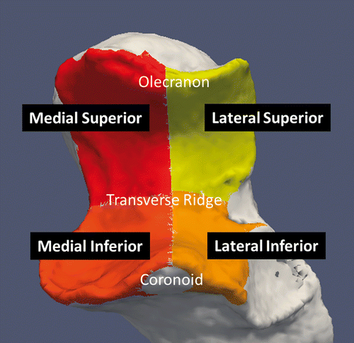
Figure 3. Registration schematic and implementation of proximity mapping. (A) Simulated elbow flexion is achieved using cadaveric specimens and the upper extremity motion simulator (shown here in the valgus gravity-dependent position). (B) Subsequent to testing, all soft tissues are removed and fiducial markers are secured for registration purposes. A second volumetric CT scan is acquired of the humerus and ulna, and homologous points are used for registration. (C) The result of this registration is a visualization of the 3D rigid-body motion of the ulna, with respect to the humerus, throughout elbow flexion. (D) Subsequent to registration of the rigid bodies, the proximity mapping technique is applied to the registered models, and the overall joint congruency can be identified for the humerus and ulna.
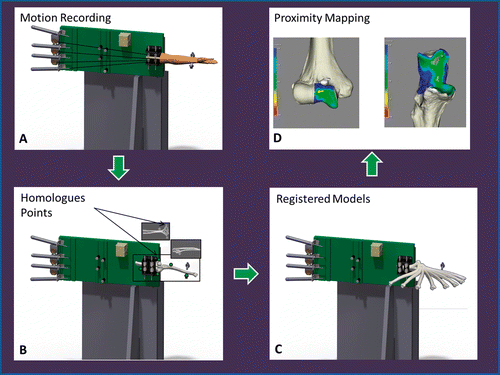
Figure 4. Proximity maps for the ulna throughout elbow flexion. Anterior view of the ulna showing the regions of close proximity (less than 4 mm).
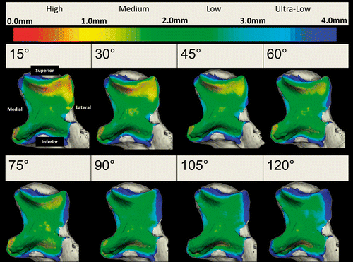
Figure 5. Surface area values for each level of proximity (high, medium, low and ultra-low) are shown throughout elbow flexion.
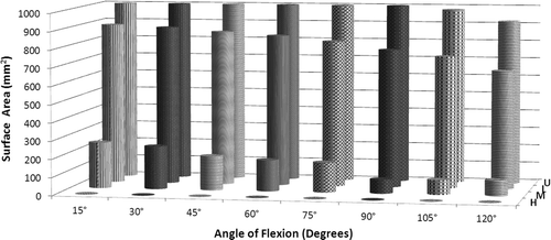
Figure 6. Surface area values for each zone at each level of proximity (high, medium, low and ultra-low) are shown as a function of elbow flexion.
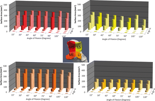
Table I. Fiducial and target registration error. Registration values are given for each specimen for the humerus and ulna, respectively.
Figure 7. Proximity mapping validation using experimental casting. The proximity maps are shown and compared to the experimental cast. The overall qualitative similarity of the cast and proximity map was assessed and used to validate the implementation of the proximity mapping technique with the registration developed in this study to examine joint surface interactions.
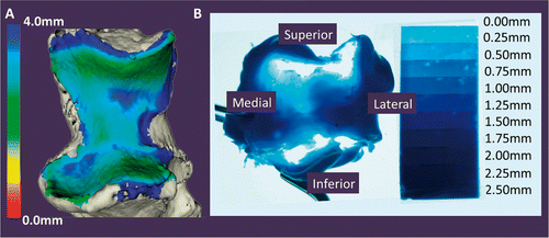
Figure 8. Comparison of experimental cast and proximity map. (A) Experimental cast. (B) The vacant regions of the cast, corresponding to regions of joint contact, were digitized using a tracked stylus. A surface model was created from this point cloud. An edge extraction filter was used to obtain the perimeter of this digitized surface. (C) The digitized cast is overlaid onto a proximity map showing the surface area on the ulna. The threshold used to generate this map is 2.87 mm, as measured from the pre-operative CT. There is 17.35% difference between the experimental cast digitization and the surface area obtained from the computational method.
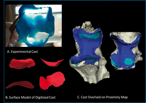
Table II. Comparison of registration error to previously developed techniques.