Figures & data
Figure 1. Structure of the device.
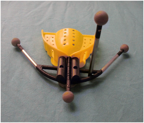
Figure 2. Preregistration of the device.
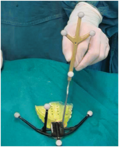
Figure 3. Measurement of 3D distances.
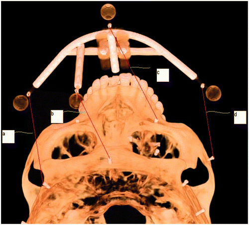
Figure 4. The skull fitted with the device.
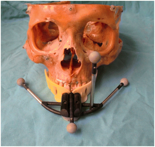
Figure 5. Comparison between the mirror of the left side (yellow) and the affected right side (blue).
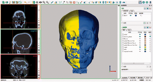
Figure 6. Pre-registration of the device before surgery.
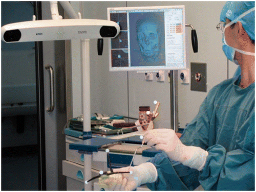
Figure 7. Reduction confirmation during the operation.
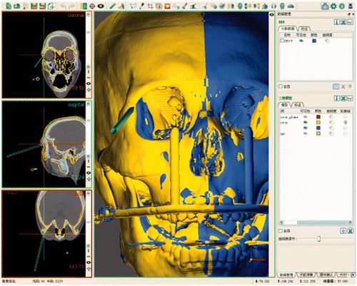
Table I. 3D distance measurements on the same skull.
Figure 8. Target fiducial color-coded visualization value (colorwash dots) of the TRE for each registration fiducial (black dots) configuration. (A) Using registration fiducials on the maxillary alveolus. (B) Using registration fiducials on the device.
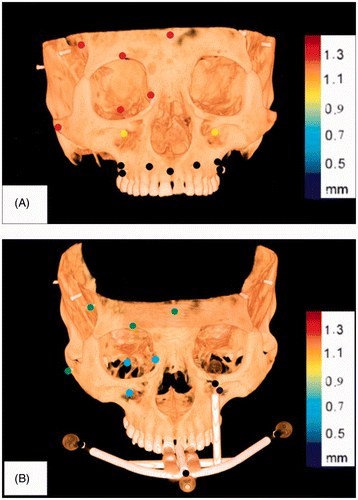
Table II. TRE of the investigated anatomical points for the fiducial configuration in which registration fiducials are defined on the maxillary alveolus and on the device.