Figures & data
Table I. Number of observations and separation into subgroups.
Figure 1. Planning of the pelvic landmarks, including the deepest point in each fossa, performed with iPlan 3.0 software (Brainlab AG).
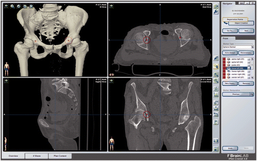
Figure 2. Alternative reference planes RP1 (ASIS-ASIS-CoR) and RP2 (ASIS-ASIS-Fossa) to define the sagittal orientation of the pelvis. The distance between the anterior rim point and the APP is also shown: This distance was used to define RP3.
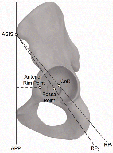
Figure 3. Measurement of the inter-teardrop distance (ITD). The measurement was performed virtually based on digitally reconstructed radiographs (DRRs) with iPlan 3.1 prototype software (Brainlab AG).
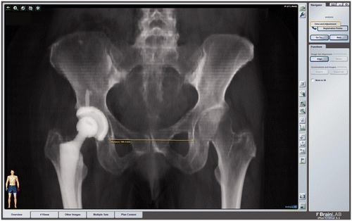
Table II. Summary of statistics (mean ± standard deviation) for anatomical relationships used in the proposed pelvis registration method.
Table III. Summary of statistics (mean ± standard deviation) for the deviation between the gold-standard APP and lateral registration, assuming a standard cup position (40° inclination, 15° anteversion).