Figures & data
Figure 1. Virtual resection planning of the cadaveric femur in the CAD software. CT images in DICOM format were imported into the CAD software. Axial (B), reformatted coronal (A) and sagittal (C) images were generated. (D) The 3D femur was segmented by the thresholding process. Planes for the bone resection (proximal TP and distal TP) were defined. The planes were 1 mm in thickness, the same as that of the oscillating saw used for osteotomy in the cadaver surgery.
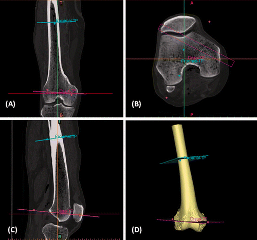
Figure 2. The 3D femur and planes of resection were exported as STL files that were then transferred to the rapid prototyping software. (A) A patient-specific CAD/CAM surgical jig was designed with cutting slits based on the exact orientation of the resection planes defined in the virtual planning. (B) The two cutting blocks were connected by a handle with an orientation that could facilitate positioning without obstructing the surgical exposure. (C) The undersurface of the surgical jig. (D) The cutting blocks had anatomical shapes that matched the bone surface at the defined resection level. This ensured easy and accurate positioning of the jig during the surgery.
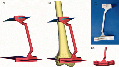
Figure 3. Preoperative screen images of surgical navigation planning on the navigation monitor for the cadaver study. The original CT image datasets (CT01) were fused with a modified CT dataset (CT02) containing the virtual CAD model of the test prosthesis, and with another modified CT dataset (CT03) containing the proximal and distal resection planes. The virtual CAD prosthesis and resection planes could be extracted with the aid of the segmentation tools in the navigation software. Proximal and distal resection planes were then marked as planned in the CAD software. Axial (A), sagittal (B) and coronal (C) images and the reconstructed 3D planning bone model (D) illustrate the transfer of the CAD virtual planning to the navigation system by the method of CAD to DICOM conversion and image fusion.
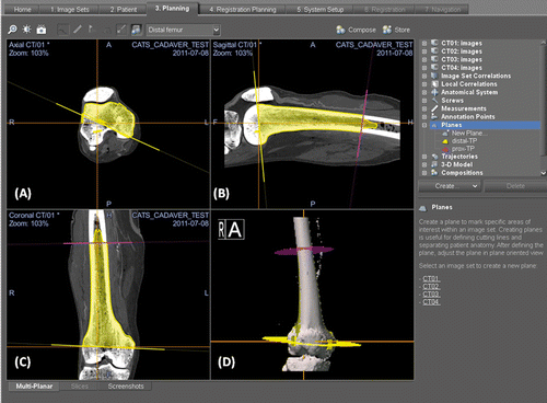
Figure 4. (A) After surgical exposure of the distal femur via a medial subvastus approach, the patient-specific CAD/CAM surgical jig was positioned at the planned resection sites. (B) The bone resection was performed with an oscillating saw through the cutting slits of the surgical template. (C) A junctional plate that matched the dimensions of the resected end during virtual simulation of the distal femoral osteotomy was compared with the distal femoral resection actually achieved. (D) The dimensional difference between the planned resection (represented by the junctional plate) and the achieved resection was <1 mm, and therefore validated the feasibility and accuracy of the technique.
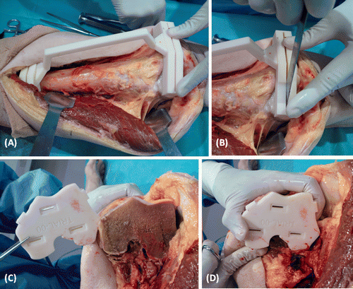
Figure 5. The accuracy of the technique was also validated with the help of a CT-based navigation system. (A) After bone resection with the aid of the surgical jig, the resected bone segment with the attached reference tracker still retained the image-to-bone registration and was used for the validation. When the navigation pointer was in contact with the resected distal bone end, the virtual tip of the pointer (the orange cross) was compared with the planned resection plane (shown in yellow) on the navigation display. The coronal (B) and sagittal (C) images and the 3D bone model (D) showed the achieved resection to differ by <1 mm from the planned resection. This suggested that the technique could help execute the preoperative planning accurately in the surgical field.
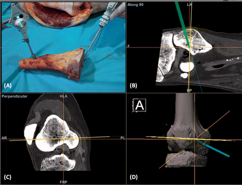
Figure 6. (A) A coronal T1-weighted MR image of a patient with a low-grade osteosarcoma of the left femur (red arrows). (B) CT images of the femur were imported into the CAD software. The surgeon defined the proximal and distal bone resections that determined the design of a CAD/CAM surgical jig. (C) The matching contour surface at the distal resection site (the green region represents the actual bone contact area). (D) A custom CAD intercalary prosthesis was designed to match exactly the bone defect after resection. Extracortical plates were added to improve the initial stability of fixation for the reconstruction.
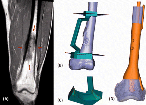
Figure 7. (A) A junctional plate was designed to validate the accuracy of the planned resection. The central slot of the plate guided the medullary position of the distal prosthetic stem. The planned location of the extracortical plate could also be marked with the junctional plate and thus helped to position the prosthesis in the correct rotational alignment. (B) The junctional plate matched well with the achieved bone resection (black arrow). (C) The distal prosthetic component fitted perfectly to the bone resection (black arrow). A cable wire was applied medially to further enhance the initial stability of the reconstruction. Cancellous bone graft was added at the bone-prosthesis junction. At two months after surgery, a plain radiograph (D) showed satisfactory alignment of the prosthesis and the patient could achieve full knee flexion (E).
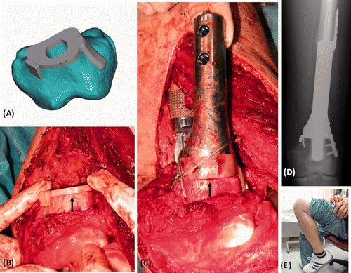
Table I. Comparison between navigation-guided and surgical jig-guided approaches for bone tumor surgery.