Figures & data
Figure 1. (a) DRF attached to the sacral hiatus with sterile tape. (b) The DRF is positioned out of the way of the surgeon.
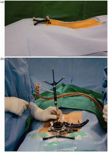
Table I. Summary of demographic data.
Figure 2. (a) Recorded navigation image and measured SAA and SAS of virtual screw location. (b) Second cb-CT scan image after screw insertion, and measured SAA of actual screw location
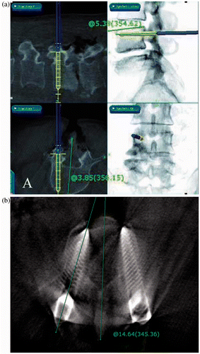
Table II. Incidence of screw breach according to fusion level.
Table III. Intraclass correlation coefficient (ICC) values in the analysis of the virtual/actual SAA and SAS validity.