Figures & data
Figure 1. T2-weighted MRI showing a right lateral soft disc herniation at C7-T1 (red arrow). Left: sagittal foraminal view; center: axial view; right: CT axial view.
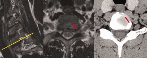
Figure 2. Operating room setting. (1) Intraoperative CT scan being performed with O-arm; (2) navigation monitor; (3) infrared camera system (IFS); (4) NIM (neural integrity monitor); and (5) dynamic reference base (DRB).
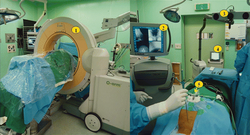
Table I. O-arm workflow.
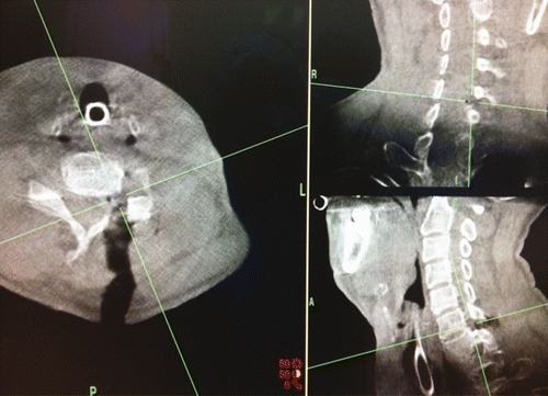
Figure 3. Left: Preoperative radiograph showing the difficulty in finding the correct level (C6-7 level) for the surgical approach. Right: O-arm navigation images allowing proper localization of the level and laminofacet junction.
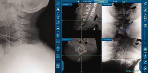
Figure 4. Left: A 1.6-cm skin incision for one-level decompression. Right: A 2.0-cm skin incision for two-level decompression.
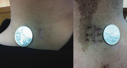
Figure 5. Postoperative posterior view of a 3D CT scan showing the small keyhole for decompression of a C6-7 left foraminal stenosis and demonstrating adequate preservation of facet joints.
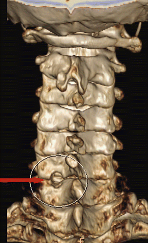
Table II. Demographics for posterior cervical microforaminotomy assisted by O-arm-based navigation.
Figure 6. Intraoperative O-arm images used to check the adequacy of decompression. A foraminal view (lower right) can be obtained to evaluate the remnant foraminal stenosis.