Figures & data
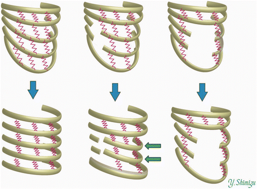
Figure 1. Mechanism of inspiration. Left: Normal inspiration. Ribs (indicated by beams) are connected with springs. As the springs contract, the ribs exhibit concerted movement. Right: When parts of the beams are removed, the movement exhibits disharmony.
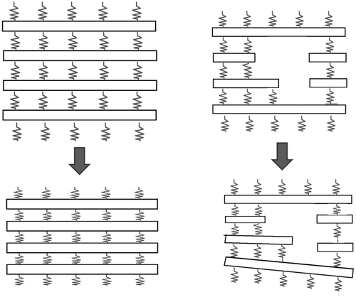
Figure 2. CT data from 16 patients (left) were transformed into 3D models consisting of beams and non-linear springs (right). See also .
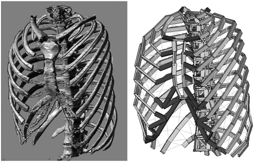
Figure 3. Inspiration muscles were modeled with non-linear springs. The red and yellow segments in the figure indicate springs simulating muscle fibers of the diaphragm and external intercostal muscles, respectively. The bones – simulated with beams – are shown thinner than in actuality to enhance the spring elements.
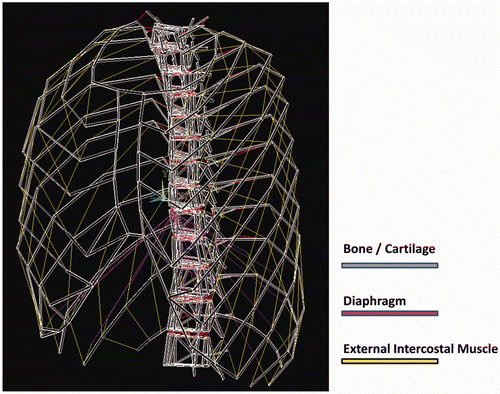
Table I. Young's moduli for bone and cartilage (MPa).
Table II. Stress-strain relationships for respiratory muscles.
Table III. Stress-strain relationships for ligaments.
Figure 4. Examples of models representing each of the harvesting patterns. Costal cartilages or ribs are harvested from the regions indicated by circles. Types II-a, II-b, and II-c are viewed from behind.
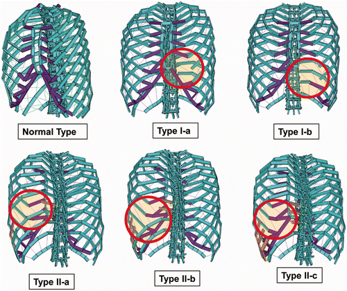
Figure 5. An example of thoracic transformation presented by a Type I-b model. The left and right figures indicate thorax shapes before (left) and after (right) inspiration. The dotted lines in the figures at right indicate the thorax shape before inspiration. The contours indicate the degree of deviation in millimeters.
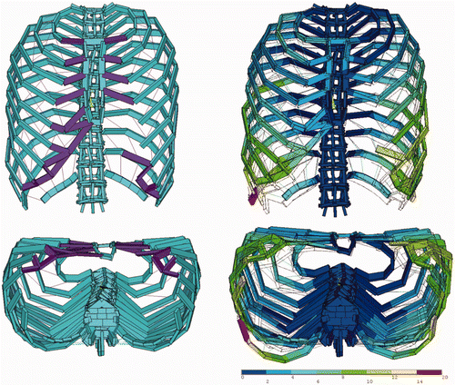
Figure 6. Upper and lower bounds of the thorax.
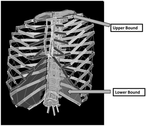
Figure 7. Position of the sternum before (left) and after (right) inspiration. θpre and θpost are the inclinations that the sternum presents relative to the coronal plane. The difference between θpost and θpre was defined as the angle of sternal rotation (ASR) and was evaluated.
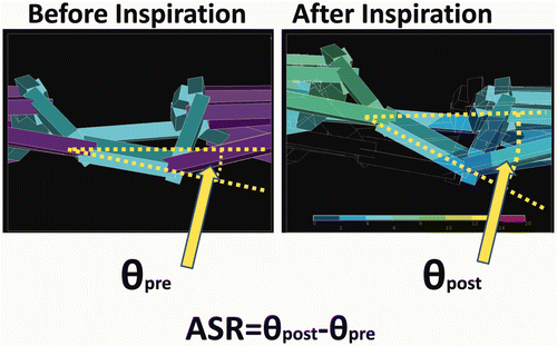
Table IV. Angle of sternal rotation (ASR).
Table V. Increase in right thoracic volume (ΔRTV).
Table VI. Increase in left thoracic volume (ΔLTV).
Table VII. Measured values as a percentage of calculated values for three volunteer subjects. Numbers in parentheses indicate the measured value (numerator) and calculated value (denominator).
Figure 8. Left: Inspiration pattern of normal thorax. With the elevation of the ribs, the volume of the thorax increases. Center: Elevation of the thorax can occur even after the removal of costal cartilages, although some ribs (indicated with arrows) exhibit deviation during inspiration. Right: Elevation of the thorax is impaired when ribs are removed from the lateral parts.