Figures & data
Figure 1. Electric actuator. The slide table position and movement speed are automatically controlled with a computer. The positioning accuracy is <0.1 mm.
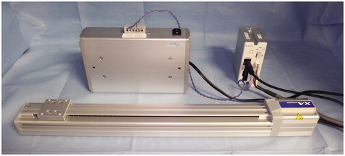
Figure 2. Range images of the maxillary first molar produced by the present system. The coordinate values of the cusp were recorded for all 30 frames, and the standard error was calculated. The left and right figures show the range images of the maxillary first molar by mesh and texture, respectively.
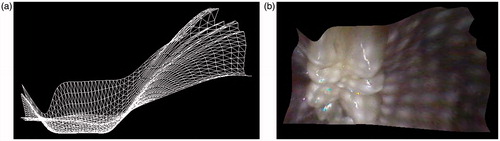
Figure 3. Fixation device comprising a commercially available light stand and a multijointed arm. This system enables arbitrary positioning.
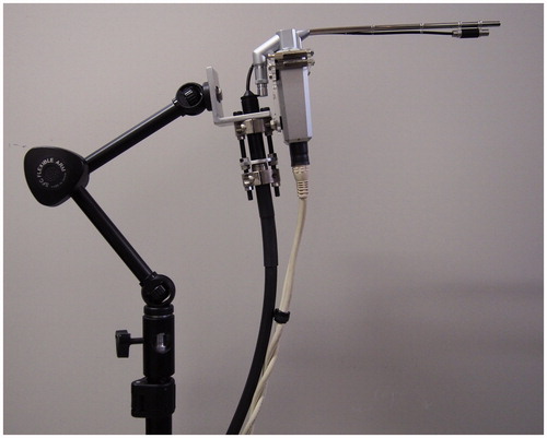
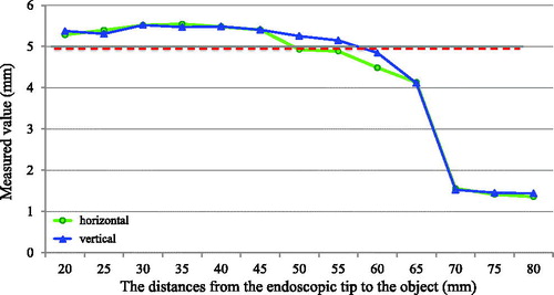
Figure 4. Relationship between the distance from the tip of the endoscope to an object and the measuring error. The measuring error became substantial when the distance from the tip of the endoscope to the object was >6.5 cm. The red dashed line shows the true distance (5 mm).