Figures & data
Figure 1. Photomicrographs of the prostatic peripheral zone from standard (a, b, c, d), HGPIN (e, f, g, h), PC (i, j, k, l) and BPH (m, n, o, p) groups. (a) Epithelium with secretory columnar and basal cells; inset: secretory epithelial cell nucleus (N), basal cells (arrow) with flattened nuclei. (b) Thin collagen fibres (Col) underlying the epithelium and among smooth muscle cells. (c), (d) Reticular (Rf) and elastic (EF) fibres underlying the epithelium. (e) HGPIN; inset: HGPIN focus, voluminous cellular nuclei (N) and basal cells (arrow). (f) Secretory epithelium with HGPIN foci (arrows), increased collagen fibres (Col) and smooth muscle cells. (g), (h) Increased reticular (Rf) and elastic (Ef) fibres. (i) Peaked neoplastic acini (asterisks) with infiltrative feature, periacinar space (arrows); Irregular smooth muscle cells; inset: neoplastic acini with increased nuclei (arrow). (j) Neoplastic acini (arrows); Increased collagen fibres (Col), smooth muscle fibres, amylaceous body (asterisk) and inflammatory cells (Ic). (k), (l) Disorganised and increased reticular (Rf) and elastic (Ef) fibres. (m) BPH with acinus showing an external straight line and internal papillomatosis. (n) Epithelium with secretory columnar and basal cells (arrow); hypercellular stroma with increased collagen fibres (Col). (o), (p) Hyper-plastic stroma with increased reticular (Rf) and elastic (Ef) fibres. a–p: Ep, epithelium, L, lumen, Smc, smooth muscle cell and St, stroma.
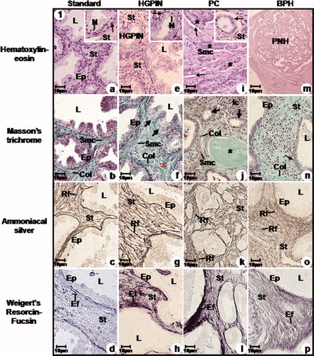
Figure 2. Electron micrographs of the prostatic peripheral zone from standard (a, b, c, d), HGPIN (e, f, g), PC (h, i, j) and BPH (k, l, m) groups. (a) Columnar cells with basal nuclei and clear nucleoli (Nu); Basal cells (Bc). Stroma (St). (b) Often secretory vesicles; short and scattered microvilli (Mv). (c) Flattened and parallel Golgi cisternae (arrow) with secretory vesicles. (d) Rough endoplasmic reticulum cisternae (RER). Basal lamina (arrow); Stroma with collagen fibres (Col) underlying the epithelium and among smooth muscle cells (Smc) and fibroblast (Fb). (e) HGPIN; Basal cells (Bc). (f) Dilated Golgi cisternae (arrow); inset: Ruptured mitochondrial cristae. (g) Dilated rough endoplasmic reticulum cisternae (RER). Digestory vacuoles (asterisk); Basal laminal (arrow); Increased collagen fibres (Col) distributed among smooth muscle cells (Smc). (h) Neoplastic cells with increased nuclei (arrow). (i) Neoplastic nuclei (arrow); amylaceous bodies (Ab); interrupted basal lamina (asterisk); Smooth muscle cells (Smc) showing irregular feature and collagen fibres (Col). (j) Neoplastic cell with increased nuclei and rare cytoplasm (arrow); Increased collagen fibres (Col). (k) Columnar cells. (l) Dilated rough endoplasmic reticulum cisternae (Rer) and Golgi (arrow). Digestory vacuoles (asterisk). (m) Basal cells (Bc); Digestory vacuoles (arrow) in the cellular cytoplasm; Increased collagen fibres (Col) and fibroblast (Fb) and smooth muscle cells (Smc); inset: smooth muscle cells showing spinous feature (Smc). a–m: L, lumen; M, mitochondria; N, nucleus; St, stroma and Vs, secretory vesicles.
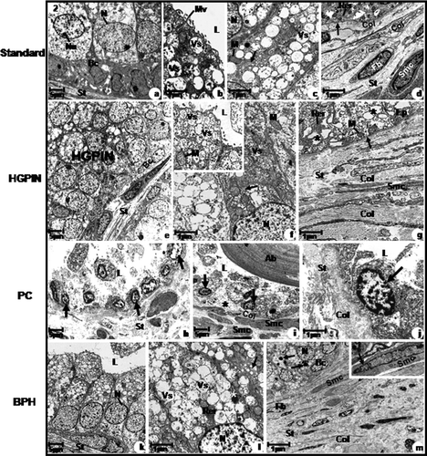
Table I. Mean and standard deviation of the nuclear, cytoplasmatic, epithelial, stromal, secretory vesicle (VS), digestory vacuoles (VD) areas (μm2) and percentage of apoptolic and proliferative index of the prostatic peripheral zone from different groups.
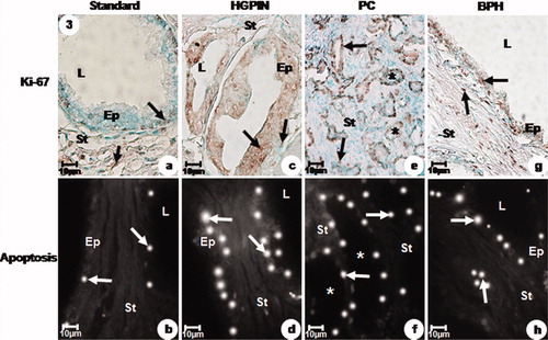
Figure 3. Immunolabelled Ki-67 and apoptotic index of the prostatic peripheral zone from standard (a, b), HGPIN (c, d), PC (e, f) and BPH (g, h) groups. (a) Weak Ki-67 immunoreactivity (arrows) in the secretory epithelial and stromal cells. (b) Weak DNA fragmentation (arrows) in the secretory epithelial cells. (c) Intense Ki-67 immunoreactivity (arrows) in the secretory epithelial and stromal cells. (d) Intense DNA fragmentation (arrows) in the secretory epithelial cells. (e) Intense Ki-67 immunoreactivity (arrows) in the neoplastic acini (asterisks) and stromal cells. (f) Moderate DNA fragmentation (arrows) in the neoplastic acini (asterisks). (g) Moderate Ki-67 immunoreactivity (arrows) in the secretory epithelial and stromal cells. (h) Moderate DNA fragmentation (arrows) in the secretory epithelial cells. a–h: Ep, epithelium, L, lumen and St, stroma.