Figures & data
Table I. Distribution by age of the elderly subjects in the TRELONG study. For a full study description see Gallucci et al. [Citation29].
Table II. Regression to assess the effect on survival of IGF-1R rs2229765 polymorphic site (a) and of circulating IL-6 (b). CVD: cardiovascular disease (including myocardial infarction, ischemic cardiopathy, peripheral vasculopathy and chronic heart failure); VC: vascular cerebral disease (including stroke and cerebral vasculopathies). C: regression coefficient; HR: hazard ratio; St. er: Standard error.
Figure 1. Survival analysis according to sex and genotype. (A) Survival for males according to IGF-1R rs2229765 (G/A) polymorphic site. (B) Survival for females according to IGF-1R rs2229765 (G/A) genotype. (C) Survival for males grouped according to IL-6 rs1800795 (C/G) polymorphic site. (D) Survival for females stratified for IL-6 rs1800795 (C/G) polymorphism.
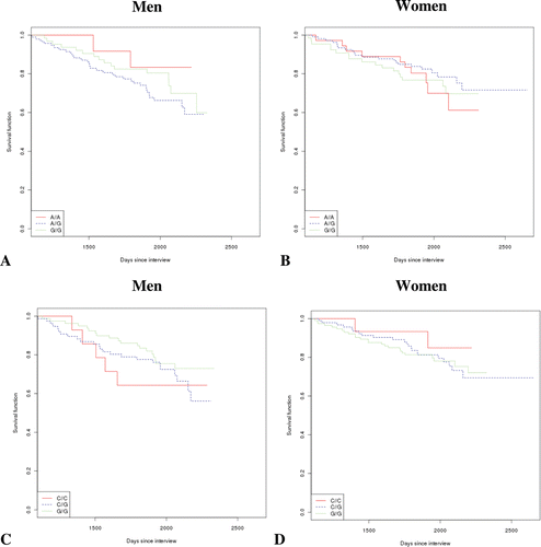
Figure 2. Survival curves on the basis of sex and circulating levels of IL-6 or IGF-1. (A) Survival in men stratified by IL-6 level in plasma (in quartiles). (B) Similar analysis in females. (C) Survival analysis according to sex and circulating levels of IGF-1 (in quartiles) in men only. No significant difference came to light considering the 1st quartile as reference and correcting for age. (D) The same analysis as in (C), for women. The individual functions showed no significant differences.
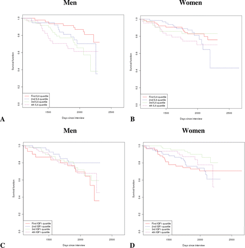
Table III. Classification of the TRELONG population according to circulating levels of IGF-1 and IL-6. p-value was calculated from Chi-square test for binary variables and ANOVA for continuous variables. CVD: cardiovascular disease (including myocardial infarction, ischemic cardiopathy, peripheral vasculopathy and chronic heart failure); VC: vascular cerebral disease (including stroke and cerebral vasculopathies); CCI: Charlson’s comorbidity index.
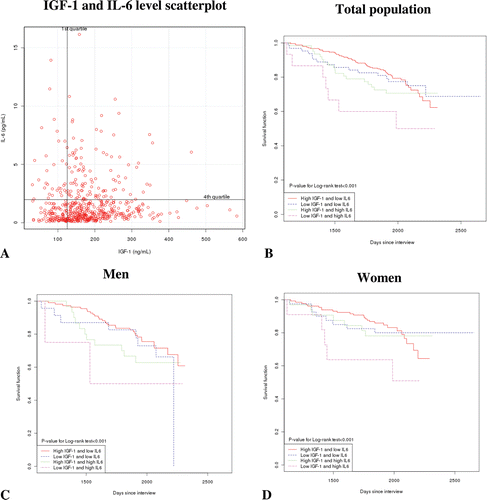
Figure 3. Assessment of combined effect of IGF-1 and IL-6 on survival. (A) The TRELONG population was divided according to low or high levels of circulating IL-6 and IGF-1 (divided into quartiles). (B) Survival curve of the entire population classified according to high/low level of IL-6 and IGF-1 (C) Survival curve as in (B) for males only; (D) Survival curve for females classified according to IL-6 and IGF-1 assessed level. For the survival analysis, the curve of high IGF-1 and low IL-6 was considered as reference.