Figures & data
Figure 1. Type I mechanoreceptor (A), Type II mechanoreceptor (B), Type III mechanoreceptor (C) and Type IV mechanoreceptor (D). S100 protein (400×).
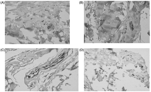
Figure 2. Correlation between age and the number of Type I (A) and Type II (B) mechanoreceptors in the femoral portion of the PCL.
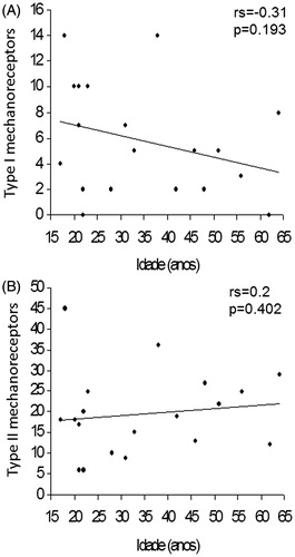
Figure 3. Correlation between age and the number of Type III (A) and Type IV (B) mechanoreceptors in the femoral portion of the PCL.
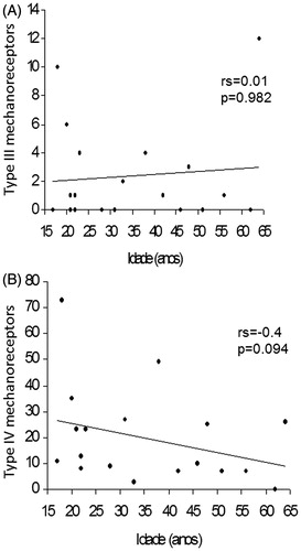
Figure 4. Correlation between age and the total number of mechanoreceptors in the femoral portion of the PCL (A) and correlation between age and the number of Type I mechanoreceptors (B).
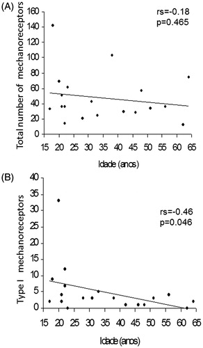
Figure 5. Correlation between age and the number of Type II (A) and Type III (B) mechanoreceptors in the tibial portion of the PCL.
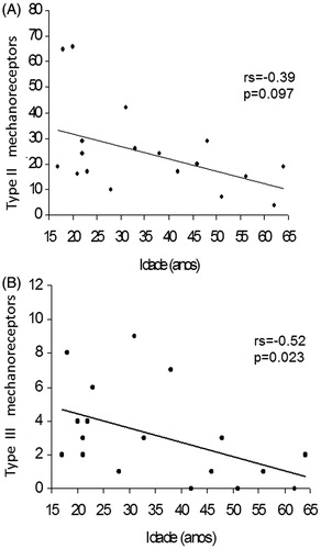
Figure 6. Correlation between age and the number of Type IV mechanoreceptors (A) and correlation between age and the total number of mechanoreceptors (B) in the tibial portion of the PCL (B).
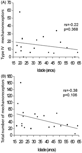
Table 1. Descriptive measures the number of Types I, II, III and IV mechanoreceptors of the femoral portion of the PCL.
Table 2. Descriptive measures regarding Types I, II, III and IV mechanoreceptors of the tibial portion of the PCL.
Table 3. Descriptive measures the total number of mechanoreceptors of the femoral and tibial portions of the PCL of 19 knees.