Figures & data
TABLE 1. Summary of mutations in CRYGS responsible for congenital cataract.
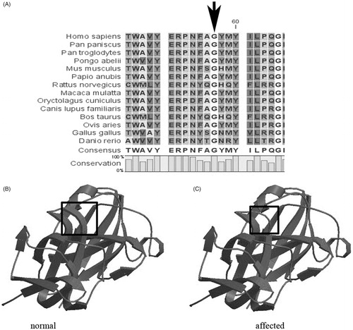
FIGURE 1. Protein models of the wild-type and mutant CRYGS. (A) A multiple-sequence alignment of the amino acid sequence in CRYGS from different species. The alignment data indicates that the Gly at the 57th amino acid position is highly conserved among many species (indicated by an arrow). (B) A structural model of the wild-type CRYGS is displayed. (C) A structural alteration of the mutant CRYGS is displayed. The second Greek key motif was found to be partially disrupted around the substitution site in the three-dimensional structural model of the mutant CRYGS.