3,152
Views
42
CrossRef citations to date
0
Altmetric
Research Article

Antioxidant, free radical-scavenging activity and cytotoxicity of different solvent extracts and their phenolic constituents from the fruit hull of mangosteen (Garcinia mangostana)

, , , , &
Pages 55-62 | Received 03 Feb 2008, Accepted 12 Nov 2008, Published online: 29 Dec 2009

Figures & data

Table 1. Contents of alpha-mangostin, total phenolic compounds, total tannin in various G. mangostana extractsa.

Table 2. IC50 of DPPH radical-scavenging activity, hydroxyl radical-scavenging, and inhibition of lipid peroxidation activity in various G.mangostana extracts and test compoundsa.

Figure 1. Effect of test compounds on H2O2-induced cell damage in keratinocyte cells when added with H2O2. Keratinocyte cells were treated with H2O2 (200 μM) with various test compounds: (A) water extract, methanol extract, and hexane extract (B) phenolic compounds contained in G. mangostana extracts (α-mangostin, epicatechin, and tannin). After 3 h of incubation, cell viability was measured using MTT assay. Data are expressed as mean ± SD (n = 3). #p < 0.05 compared with the H2O2-treated group.
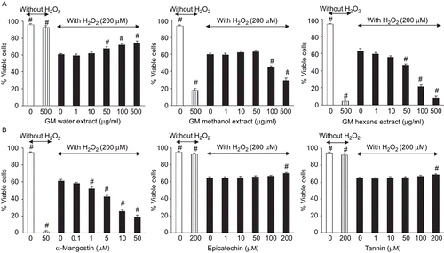
Figure 1.  Effect of test compounds on H2O2-induced cell damage in keratinocyte cells when added with H2O2. Keratinocyte cells were treated with H2O2 (200 μM) with various test compounds: (A) water extract, methanol extract, and hexane extract (B) phenolic compounds contained in G. mangostana extracts (α-mangostin, epicatechin, and tannin). After 3 h of incubation, cell viability was measured using MTT assay. Data are expressed as mean ± SD (n = 3). #p < 0.05 compared with the H2O2-treated group.

Figure 2. Effect of test compounds on toxicity in keratinocyte cells. Keratinocyte cells were treated with various test compounds: (A) methanol extract, (B) hexane extract, (C) α-mangostin. After 24 h of incubation, cell viability was measured using MTT assay. Data are expressed as mean ± SD (n = 3). #p < 0.05 compared with the control (without test compounds).
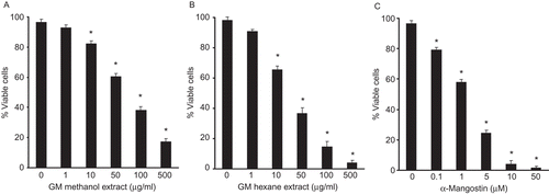
Figure 2.  Effect of test compounds on toxicity in keratinocyte cells. Keratinocyte cells were treated with various test compounds: (A) methanol extract, (B) hexane extract, (C) α-mangostin. After 24 h of incubation, cell viability was measured using MTT assay. Data are expressed as mean ± SD (n = 3). #p < 0.05 compared with the control (without test compounds).

Reprints and Corporate Permissions

Please note: Selecting permissions does not provide access to the full text of the article, please see our help page How do I view content?

To request a reprint or corporate permissions for this article, please click on the relevant link below:

Academic Permissions

Please note: Selecting permissions does not provide access to the full text of the article, please see our help page How do I view content?

Obtain permissions instantly via Rightslink by clicking on the button below:

If you are unable to obtain permissions via Rightslink, please complete and submit this Permissions form. For more information, please visit our Permissions help page.