997
Views
7
CrossRef citations to date
0
Altmetric
Research Article

Hemagglutinating activity of proteins from Parkia speciosa seeds

, , &
Pages 81-88 | Received 31 Jul 2008, Accepted 08 Nov 2008, Published online: 29 Dec 2009

Figures & data

Table 1. Hemagglutinating activities and yields at various stages of purification of lectin from Parkia speciosa.

Figure 1. Affinity chromatogram of crude protein of Parkia speciosa on Affi-Gel Blue gel column (1.6 × 10 cm) in binding buffer 1 mM Tris-HCl, pH 7.2. Dashed lines indicate use of 0–100% linear gradient of 0.5 mM NaCl in binding buffer. Flow rate 1.5 mL/min.
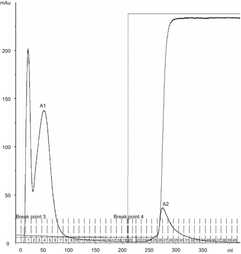
Figure 1.  Affinity chromatogram of crude protein of Parkia speciosa on Affi-Gel Blue gel column (1.6 × 10 cm) in binding buffer 1 mM Tris-HCl, pH 7.2. Dashed lines indicate use of 0–100% linear gradient of 0.5 mM NaCl in binding buffer. Flow rate 1.5 mL/min.

Figure 2. Gel filtration chromatogram of A1, A2 fractions from Affi-Gel Blue gel chromatography on Superdex 200 column (1.6 × 60 cm) in double distilled water. Flow rate 0.5 mL/min.
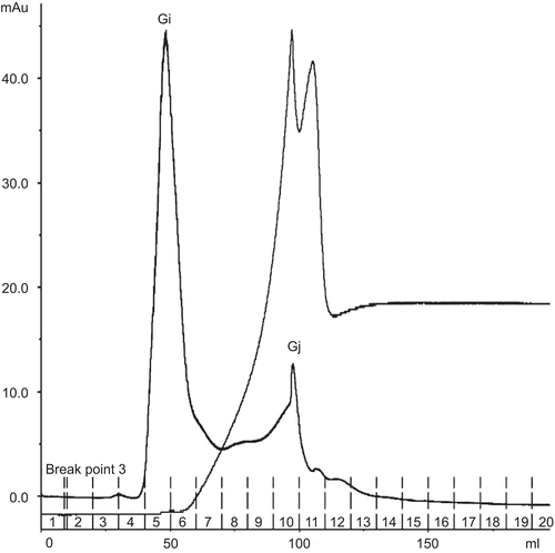
Figure 2.  Gel filtration chromatogram of A1, A2 fractions from Affi-Gel Blue gel chromatography on Superdex 200 column (1.6 × 60 cm) in double distilled water. Flow rate 0.5 mL/min.

Figure 3. MALDI-ToF mass spectrum of Gj fraction from Superdex 200 gel filtration.

Figure 3.  MALDI-ToF mass spectrum of Gj fraction from Superdex 200 gel filtration.

Table 2. Proteins identified by MALDI-ToF mass spectrometry followed by peptide mass fingerprinting via the Mascot search engine.

Figure 4. Affinity chromatogram of crude protein from Parkia speciosa on Con A Sepharose column (1.6 × 5 cm). Flow rate 1.5 mL/min. The equilibration buffer was 20 mM Tris-HCl, pH 7.4, containing 0.5 M NaCl, 1 mM CaCl2·2H2O, 1 mM MnCl2. The eluted lectin buffer was 20 mM Tris-HCl, pH 7.4, containing 0.1–0.5 M methyl-α-d-mannopyranoside.
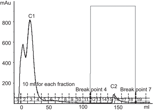
Figure 4.  Affinity chromatogram of crude protein from Parkia speciosa on Con A Sepharose column (1.6 × 5 cm). Flow rate 1.5 mL/min. The equilibration buffer was 20 mM Tris-HCl, pH 7.4, containing 0.5 M NaCl, 1 mM CaCl2·2H2O, 1 mM MnCl2. The eluted lectin buffer was 20 mM Tris-HCl, pH 7.4, containing 0.1–0.5 M methyl-α-d-mannopyranoside.
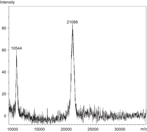
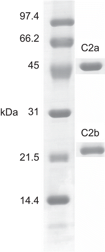
Figure 5. One-dimensional SDS-PAGE of lectin from Parkia speciosa (C2a and C2b) was visualized with Coomassie staining. 12.5% acrylamide gel was used.

Figure 5.  One-dimensional SDS-PAGE of lectin from Parkia speciosa (C2a and C2b) was visualized with Coomassie staining. 12.5% acrylamide gel was used.

Reprints and Corporate Permissions

Please note: Selecting permissions does not provide access to the full text of the article, please see our help page How do I view content?

To request a reprint or corporate permissions for this article, please click on the relevant link below:

Academic Permissions

Please note: Selecting permissions does not provide access to the full text of the article, please see our help page How do I view content?

Obtain permissions instantly via Rightslink by clicking on the button below:

If you are unable to obtain permissions via Rightslink, please complete and submit this Permissions form. For more information, please visit our Permissions help page.