Figures & data
Table 1. Composition of Echinacea extracts.
Figure 1. Effect of aqueous Echinacea extract EP-3 on the extracellular proliferation of L. donovani. Exponentially growing L. donovani were incubated for 24, 48 and 72 h with the indicated dilutions of Echinacea extract EP-3. Control Leishmania were grown under identical conditions without Echinacea. At each time point the numbers of motile surviving parasites were counted by Trypan blue exclusion. The data shown are representative of three independent experiments: lower black bars, 24 h; grey bars, 48 h; upper white bars, 72 h.
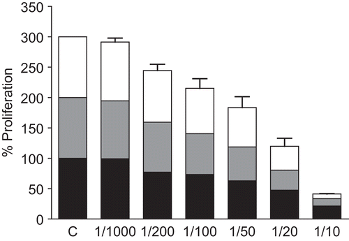
Figure 2. Effect of ethanol Echinacea extract EA on the extracellular proliferation of L. donovani. Exponentially growing L. donovani were incubated for 24, 48 and 72 h with the indicated dilutions of EA (left hand bars; lower black bars, 24 h; middle grey bars, 48 h; upper white bars, 72 h). Controls consisted of dilutions of ethanol corresponding to those in the diluted extracts (right hand bars; lower bars, 24 h; middle bars, 48 h; upper bars, 72 h). At each time point the numbers of motile surviving parasites was counted by Trypan blue exclusion. The data shown are representative of three independent experiments.
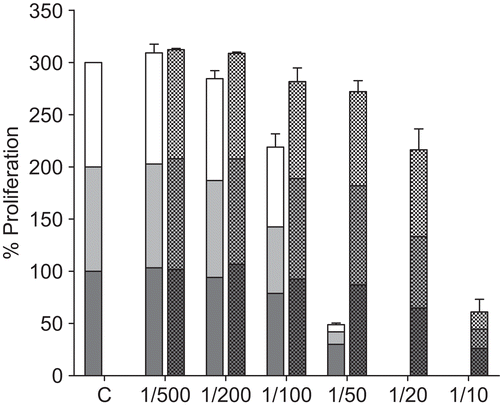
Figure 3. Effect of aqueous Echinacea extract EP-3 on the extracellular proliferation of T. brucei. Exponentially growing T. brucei were incubated for 24, 48 and 72 h with the indicated dilutions of Echinacea extract EP-3. Control trypanosomes were grown under identical conditions without Echinacea. At each time point the numbers of motile surviving parasites was counted by Trypan blue exclusion. The data shown are representative of three independent experiments: lower black bars, 24 h; grey bars, 48 h; upper white bars, 72 h.
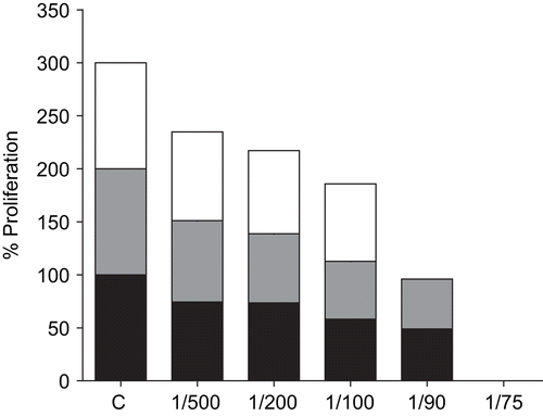
Figure 4. Effect of ethanol Echinacea extract EA on the extracellular proliferation of T. brucei. Exponentially growing T. brucei were incubated for 24, 48 and 72 h with the indicated dilutions of Echinacea extract EA (left hand bars; lower black bars, 24 h; middle grey bars, 48 h; upper white bars, 72 h). Controls consisted of dilutions of ethanol corresponding to the diluted extracts (right hand bars; lower bars, 24 h; middle bars, 48 h; upper bars, 72 h). At the end of the experiment, the number of motile surviving parasites was counted by Trypan blue exclusion. The data shown are representative of three independent experiments.
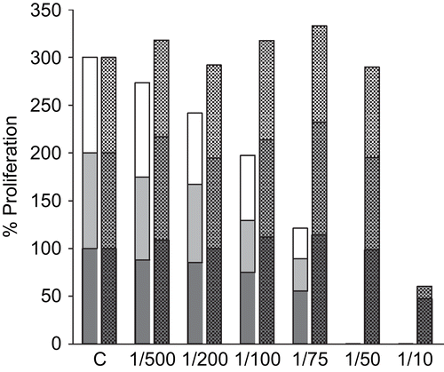
Table 2. Effect of Echinacea extract EP-1 on induced secretion of pro-inflammatory cytokines.