Figures & data
Figure 1. Effect of GMDZ decoction on the scores of ambulation (A) and rearing (B) on day 33 in the open field test. The changes of ambulation before and during the UCMS protocol (C), and the changes of rearing before and during the UCMS protocol (D). The stress was initiated at day 0. No significant difference between animals in the four groups was present at day 0 (base line).Each column represents the mean ± SEM of 9–11 animals. +p <0.05; ++p <0.01 compared with UCMS group.*p <0.05; **p <0.01 compared with the vehicle-treated control.
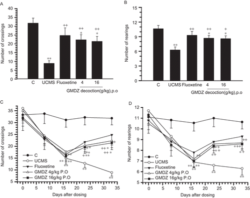
Table 1. The sucrose preference test before unpredicted chronic mild stress (![]() − SE).
− SE).
Table 2. Effects of GMDZ decoction in sucrose preference test in UCMS treated rats (![]() − SE).
− SE).
Figure 2. Effect of GMDZ decoction on glutamate levels in the frontal cortex and hippocampus. (A) Typical chromatograms of amino acid standard; concentrations of amino acids in the standard solution are 0.6 μg/mL. (B) Chromatograms of rat frontal cortex and hippocampus sample. (C) Glutamate levels in the hippocampus. (D) Glutamate levels in the frontal cortex. The measured values for glutamate level in brain regions were expressed in μg/g wet tissue weight. Each column represents the mean ± SEM of 9–11 animals. +p <0.05; + +p <0.01 compared with UCMS group;*p <0.05; **p <0.01 compared with the vehicle-treated control; Glu, glutamate.
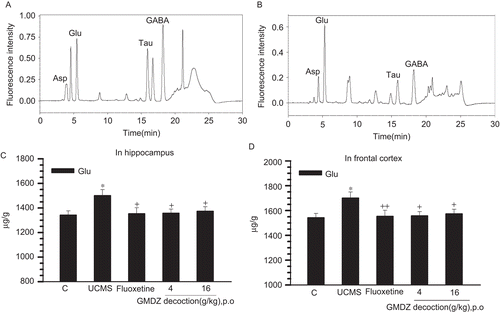
Figure 3. Western blots were carried out to analyze the expression of NMDA receptor subunits (NR1; NR2A; NR2B) and β-actin in crude membranes isolated from rat frontal cortex (A) and hippocampus (B) after administration of GMDZ decoction. Results shown are immunoblots from single representative experiments. (1) Control group, (2) UCMS group, (3) UCMS + fluoxetine, (4) UCMS + low-dose GMDZ decoction, (5) UCMS + high-dose GMDZ decoction.
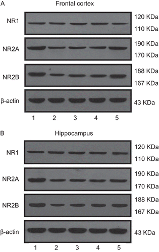
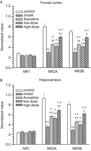
Figure 4. Expression of NMDA receptor subunits in crude membranes isolated from rat frontal cortex (A) and hippocampus (B) after administration of GMDZ decoction. Western blots were used to analyze the expression of NMDA receptor subunits. The relative expression values were normalized with β-actin value. Data are presented as mean ± SEM; +p <0.05; ++p<0.01 compared with UCMS group;*p <0.05; **p <0.01 compared with the vehicle-treated control.