Figures & data
Figure 1. Chemical structure of acetylshikonin (1), and β,β-dimethylacrylshikonin (2).
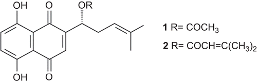
Table 1. Sequence of primers.
Figure 2. Inhibitory effect of Aikete injection on SMMC-7721. (A) SMMC-7721 cells were treated with various concentrations of Aikete injection for 48 h, then tested with MTT; (B) the percentage of cell viability was determined by Trypan blue dye assay after 1, 2, 3, 4, 5, 6, and 7 days of treatment, respectively. Data were the mean ± SD of three independent experiments.
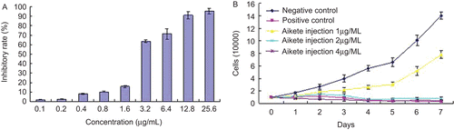
Figure 3. Morphological changes of SMMC-7721 cells detected by Giemsa staining (×200). SMMC-7721 cells incubated in the absence (A) or presence (B) of 2.0 µg/mL Aikete injection for 48 h were stained with Giemsa. Cells were viewed under microscope.
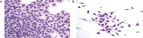
Figure 4. Morphological changes of SMMC-7721 cells detected by Hoechst 33258 staining (×400). After untreated (A) and treated with 1.0 µg/mL (B), 2.0 µg/mL (C), 4.0 µg/mL (D) Aikete injection for 48 h, cells were stained with Hoechst 33258 and observed under fluorescence microscope. Arrows indicate apoptotic cells.
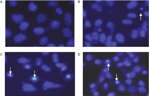
Table 2. Effect of Aikete injection on apoptosis rates and cell cycle of SMMC-7721 cells (n = 3, mean ± SD).
Figure 5. Effect of Aikete injection on the expression of Bcl-2/Bax in SMMC-7721 cells. Different concentrations of Aikete injection were added to SMMC-7721 cell culture for 48 h and then cells were harvest to be processed. The expression of Bcl-2 and Bax were analyzed by flow cytometry. Data were the mean ± SD of three independent experiments. Compare with negative control: *P < 0.05 **P < 0.01
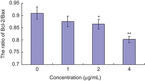
Figure 6. Effect of Aikete injection on Bcl-2 and Bax mRNA expression in SMMC-7721 cells. Cells were treated with various concentrations of Aikete injection for 48 h. The mRNA levels of Bcl-2 and Bax were analyzed by RT-PCR, and actin was used as a control. Data were the mean ± SD of three independent experiments. Compare with negative control: *P < 0.05 **P < 0.01.
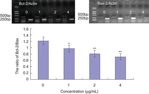
Table 3. Effect of Aikete injection on H22 bearing mice (mean ± SD).
Figure 7. Anti-tumor effects of Aikete injection in vivo. (A) Growth curve of mouse transplantable hepatoma; (B) inhibitory effects of Aikete injection and cyclophosphamide on mouse tumor. Transplanted tumors from each group were shown: (B1) negative control; (B2) solvent control; (B3) cyclophosphamide 60 mg/kg; (B4) Aikete injection 0.5 mg/kg; (B5) Aikete injection 1.0 mg/kg; (B6) Aikete injection 2.0 mg/kg. Data were the mean ± SD of three independent experiments.
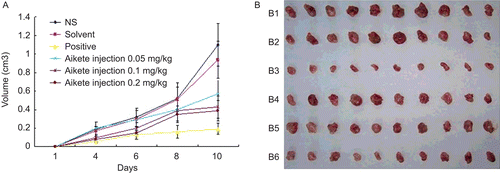
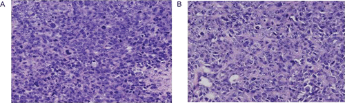
Figure 8. Morphological changes of H22 tumor cells treated with HE staining (×200). The transplanted tumors in the group of negative control (A) and Aikete injection 2.0 mg/kg (B) were exercised for HE staining and observed under microscope.