Figures & data
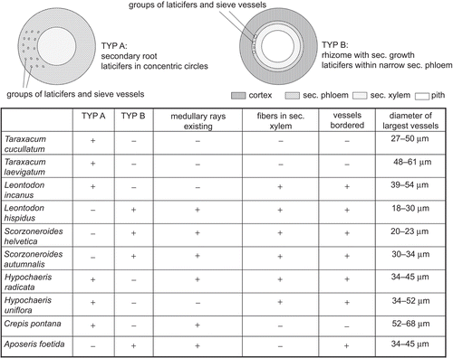
Table 1. List of species examined in this study (plants arranged according to the classification applied by CitationFischer et al., 2008).
Figure 1. Taproot of (A) T. cucullatum and (B) T. laevigatum. (C) Schematic view of Taraxacum sp. secondary root in transverse section: small ellipses mark the position of the laticifers within the dominating secondary phloem (gray), secondary xylem with vessels dispersed; medullary rays are not found.
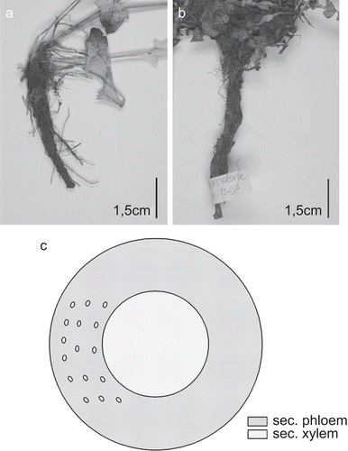
Figure 2. Taraxacum roots. (A) Overview showing the extension and arrangement of the diverse tissues: cortex lost due to formation of rhytidome, secondary phloem dominating, laticiferous vessels within secondary phloem circularly arranged, medullary rays in secondary xylem missing; (B) secondary phloem with laticiferous vessels arranged in concentric circles; (C) secondary xylem with vessels irregularly dispersed over transverse section, fibers missing; (D) reticulate vessels of secondary xylem [(A, C) T. laevigatum (TL01-08), (B, D) T. cucullatum (TC03-08)]; (A–C) transverse sections; (D) longitudinal section; scale bars = 50 µm.
![Figure 2. Taraxacum roots. (A) Overview showing the extension and arrangement of the diverse tissues: cortex lost due to formation of rhytidome, secondary phloem dominating, laticiferous vessels within secondary phloem circularly arranged, medullary rays in secondary xylem missing; (B) secondary phloem with laticiferous vessels arranged in concentric circles; (C) secondary xylem with vessels irregularly dispersed over transverse section, fibers missing; (D) reticulate vessels of secondary xylem [(A, C) T. laevigatum (TL01-08), (B, D) T. cucullatum (TC03-08)]; (A–C) transverse sections; (D) longitudinal section; scale bars = 50 µm.](/cms/asset/32547c91-0c15-4915-9b0c-cacb54cc95af/iphb_a_548390_f0002_b.gif)
Figure 4. (A) Rhizome of S. hispanica. (B) Schematic section of rhizome: small ellipses mark the position of the laticifers; vascular cylinder with vessels (dots) arranged in single or multiple radial rows.
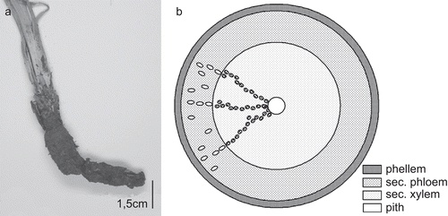
Figure 5. S. hispanica rhizome. (A, B) Overview showing the extension and arrangement of tissues: secondary phloem with laticiferous vessels arranged in rows, fibers are missing (A), secondary xylem dominated by parenchymatous cells with fibers missing, vessels arranged in single to multiple rows (B) (SHI03); (C) phellem thin-walled, thoroughly parallel-laminated (SHI02); (D) vessels of secondary xylem more or less in single or double rows (SHI02); (A–D) transverse sections; scale bars = 50 µm.
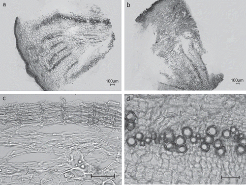
Figure 6. (A) Taproot of C. intybus. (B) Schematic view of C. intybus root in transverse section: secondary root: small ellipses mark the laticifers, dots within xylem are vessels.
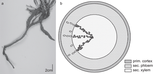
Figure 7. C. intybus root. (A) Overview showing the extension and arrangement of tissues: secondary phloem and secondary xylem are of similar extension (S5); (B) secondary phloem with lactiferous vessels arranged in rows (CI1-08); (C) secondary xylem with vessels in groups and short rows (CI1-08); (D) secondary xylem: vessels and rays (CI2-07); (A–C) transverse sections; (D) longitudinal section; scale bars = 50 µm.
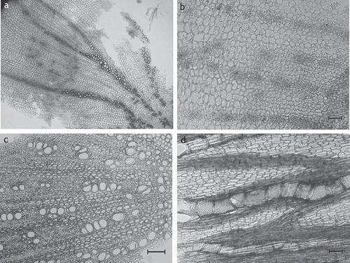
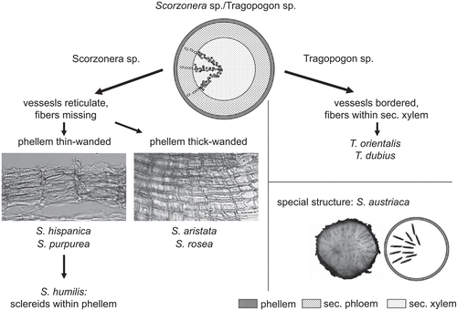
Figure 8. Differentiation of Scorzonera sp. and Tragopogon sp.; S. austriaca: transverse section treated with phloroglucine-HCL, lignified tracheids of the xylem parts red-colored.
