Figures & data
Figure 1. HPTLC Fingerprint of aqueous extract of roots of Boerhaavia diffusa Linn.
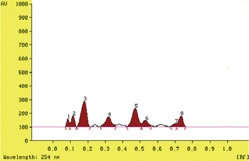
Figure 2. DPPH free radicals scavenging activity BDE with reference to standard (ascorbic acid).
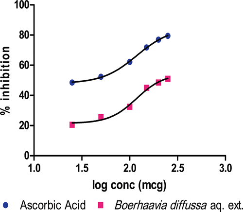
Table 1. Effect of BDE treatment on oxalate excretion and renal function in rats.
Figure 3. CaOx crystals in the urine of different treatment groups (Leica EZ-4D) at 40 × 10× magnification. (A) Control rats showed very few or no crystals; (B) Untreated rats excreted numerous oval COM and pyramidal shaped COD crystals; (C and D) Treated rats excreted a significantly reduced number of crystals.
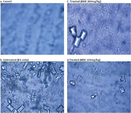
Table 2. Effect of BDE treatment on renal enzymes and other antioxidant markers in rat kidney homogenate analysis.
Table 3. Histopathological analysis of hyperoxaluria-induced renal damage and crystal deposits.
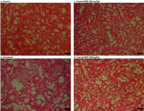
Figure 4. Histopathological examination of kidney slides of different treatment groups (Leica EZ-4D) at 10 × 10× magnification. (A) Control rats showing normal renal architecture; (B) Kidney of untreated rats showing polymorphic irregular crystal deposits tubular damage (in figure, black spots inside the rectangular box are crystal deposits); (C) BDE-treated (100 mg/kg) showed few crystal deposits and little dilation of tubules; (D) BDE-treated (200 mg/kg) showed a very few or no crystal deposits and nearly normal renal architecture.