Figures & data
Figure 1. Influence of ginkgo-derived CMC ethanol extract sample treatment on the viability of the RAW 264.7 macrophage cell line. RAW 264.7 cells were pre-treated with various concentrations of each sample before adding 1 μg/mL LPS. After 24 h of incubation, the cells were processed using MTT assay to determine viability of the cells. This figure shows the results obtained after treating 50 μg/mL of each sample. CE represents the crude initial ethanol extract of the ginkgo-derived CMCs before fractionation. **p < 0.01 indicates significant differences.
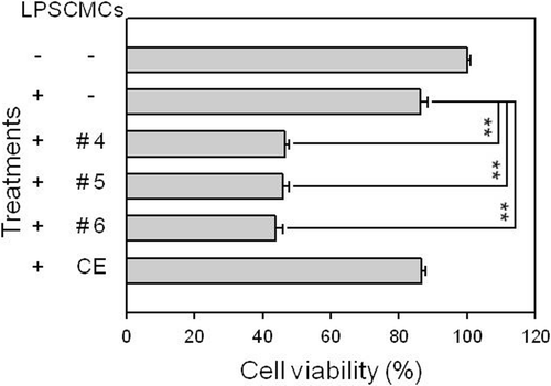
Figure 2. Effect of fractionated sample treatments on nitrite production in the LPS-stimulated RAW 264.7 macrophage cell line. RAW 264.7 cells were pre-treated with various concentrations of each sample before adding 1 μg/mL LPS. After 24 h of incubation, the culture supernatant of the cells was processed to measure nitrite production. This figure shows the results obtained after treating 50 μg/mL of each sample. CE represents the crude initial ethanol extract of the ginkgo-derived CMCs before fractionation. *p < 0.05, **p < 0.01, and ***p < 0.001 indicate significant differences.
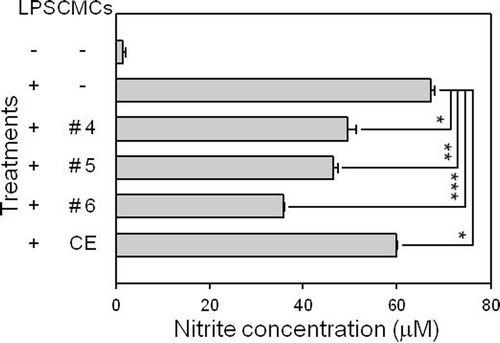
Figure 3. Influence of fractionated sample treatment on LPS-induced iNOS and COX-2 gene transcription in RAW 264.7 cells. RAW 264.7 cells were pre-treated with various concentrations of each sample before adding 1 μg/mL LPS. After 24 h of incubation, total RNA was extracted and RT-PCR was performed. Levels of GAPDH gene, a RT-PCR product, were used as an internal control. This figure shows the results obtained after treating 50 μg/mL of each sample. CE represents the crude initial ethanol extract of the ginkgo-derived CMCs before fractionation.
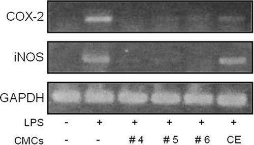
Figure 4. Influence of fractionated sample treatment on phosphorylation of ERK and p38 MAP kinases in LPS-stimulated RAW 264.7 cells. Cells were exposed to 50 μg/mL of the fractionated samples 30 min before exposure to LPS (1 μg/mL). Thirty minutes after the LPS treatment, phosphorylated ERK and p38 levels were detected using specific antibodies. The level of β-actin protein expression was used as an internal control. CE represents the crude ethanol extract, which was the initial ethanol extract prepared from ginkgo-derived CMCs before fractionation.
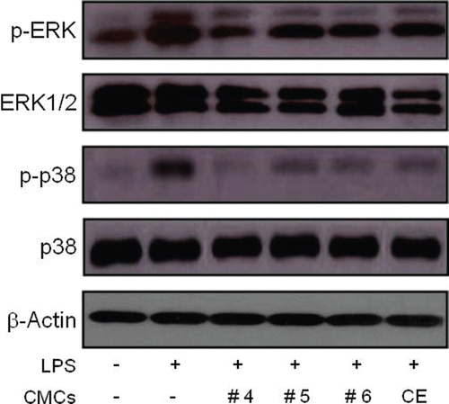
Figure 5. Influence of fractionated sample treatment on the activation of NF-κB and phosphorylation of IκB in LPS-stimulated RAW 264.7 cells. Cells were exposed to 50 μg/mL of the fractionated samples 30 min prior to the LPS (1 μg/mL) treatment. Two hours after the LPS treatment, nuclear extract and whole cell lysates were extracted and NF-κB and IκB levels were detected using specific antibodies from the nuclear extract and whole cell lysate, respectively. HDAC2 protein levels were used as an internal control. CE represents the crude ethanol extract, which was the initial ethanol extract of the ginkgo-derived CMCs before fractionation.
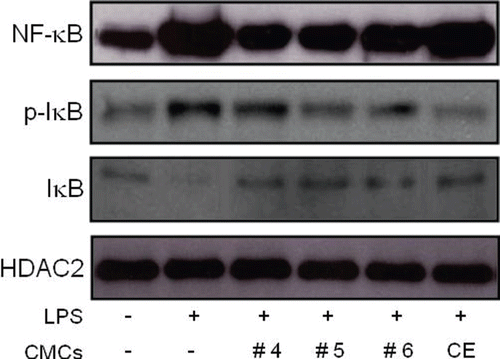
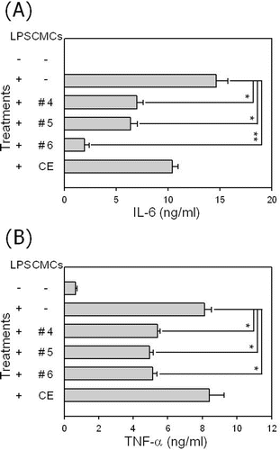
Figure 6. Influence of fractionated sample treatment on the production of pro-inflammatory cytokines in LPS-stimulated RAW 264.7 cells. Cells were stimulated with 1 μg/mL of LPS in the presence of a 50 μg/mL fractionated sample treatment. After 48 h of incubation pro-inflammatory cytokines TNF-α, and IL-6 were measured in the culture supernatant using ELISA. Data represent the mean value of triplicates and *p < 0.05 and **p < 0.01 indicate significant differences.