Figures & data
Figure 1. . Structure of UR solic acid.
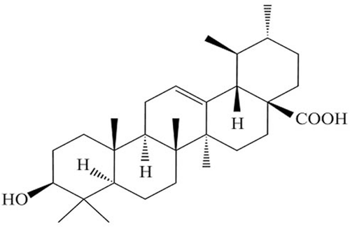
Figure 2. Stimulatory effects of UA on liver mass recovery after 70% PHx. liver to body weight ratio was examined in the livers at pre PHx, and at 0, 24, 36, 48, 72, 168 and 336 h after PHx. Data are expressed as means ± SD. n = 5 in each group. *P < 0.05 versus sham group; #P < 0.05 versus vehicle-treated control group. UA, ursolic acid.
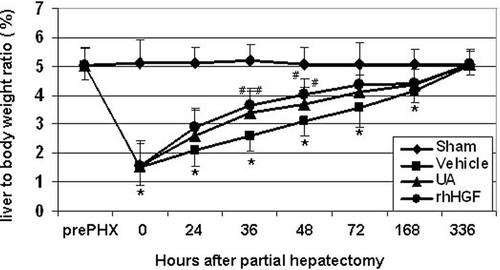
Figure 3. Hepatocyte proliferation enhancement by UA following 70% PHx (A) Representative sections of BrdU labeling of hepatocytes nuclei in the sham, control, UA and HGF treated mice at 36 h following PHx. (Magnification, × 200). (B) Summary of BrdU labeling index. Values are means ± SD (n = 5). *P < 0.05; **P < 0.01 versus sham group; #P < 0.05, # #P < 0.01 versus vehicle-treated control group.
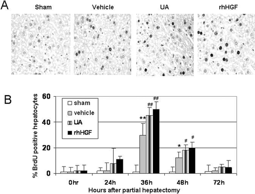
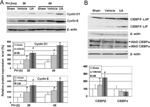
Figure 4. Western blotting for cell cycle proteins and C/EBPβ expression. (A) Expression of cyclin D1 and cyclin E proteins were increased at 36 and 48 h after PHx in UA treated group. (B) Expression of C/EBPβ proteins was increased at 36 h after PHx in UA treated group animals. Changes in the protein levels relative to control were assessed by scanning densitometry of the immunoblots. Data represent mean ± SD (n = 3). *P < 0.05 versus sham group; #P < 0.05 versus vehicle-treated control group.