Figures & data
Figure 1. Chromatographic separation of pDNA isoforms with HIC. Linear gradient was performed at 400 cm/h by decreasing the ammonium sulfate concentration. (A) 2–1 M ammonium sulfate in 40 mM Tris/HCl, pH 8.0, (B) 2.5–1 M ammonium sulfate in 40 mM Tris/HCl, pH 8.0, (C) 3–1 M ammonium sulfate in 40 mM Tris/HCl, pH 8.0.
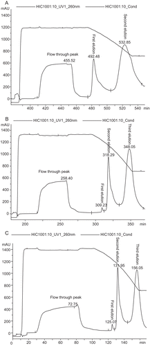
Figure 2. Agarose gel electrophoresis analysis. Lane 1: lysate; Lane 2, 6, 12: loading sample; Lane 3, 7, 13: Flow-through peak; Lane 4, 8, 14: First elution peak; Lane 5, 9, 15: Second elution peak; Lane 10, 16: Third elution peak; Lane 17: DNA ladder.
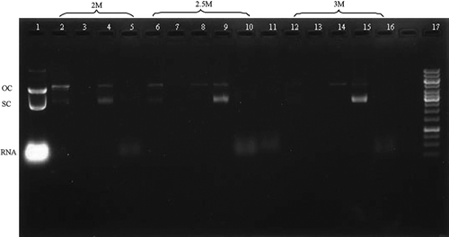
Figure 3. Plasmid purity analysis by HPLC. Plasmid purity was determined by anion-exchange HPLC on a MONO QTM 5/50 GL column. A linear gradient was performed at 0.5 mL/min by increasing the NaCl 0–1 M in 40 mM Tris/HCl, pH 8.0 for 10 CV at a flow rate of 0.5 mL/min. (A) plasmid sample purified by 2 M ammonium sulfate, (B) plasmid sample purified by 2.5 M ammonium sulfate, (C) plasmid sample purified by 3 M ammonium sulfate.
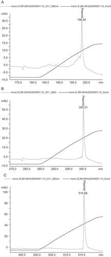
Figure 4. Result of inverted fluorescence microscope (×100). A: Plasmid purified using OMEGA kit B: Plasmid purified by 3 M ammonium sulfate.
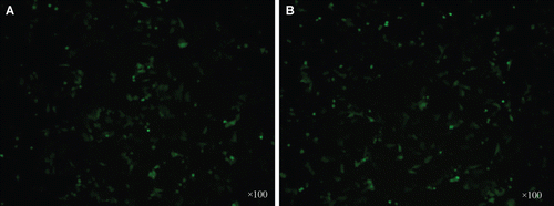
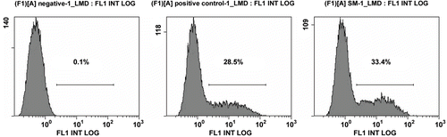
Figure 5. Transfection efficiency detected by flow cytometry. (A) negative (B) Plasmid purified using OMEGA kit C: Plasmid purified by 3 M ammonium sulfate.