2,712
Views
27
CrossRef citations to date
0
Altmetric
Research Article

The effect of silymarin (Silybum marianum) on human skin fibroblasts in an in vitro wound healing model

, , , , , , , , , & show all
Pages 298-303 | Received 20 May 2012, Accepted 14 Aug 2012, Published online: 08 Nov 2012

Figures & data

Figure 1.  Effect of silymarin on cellular viability and proliferation of human fibroblast cells. Cells were treated with different concentrations of silymarin for 24 and 48 h. (A) Cell viability was determined by the MTT assay. (B) Cell proliferation was determined by DNA (BrdU) incorporation assay. The data are expressed as mean ± SE from at least three separate experiments.
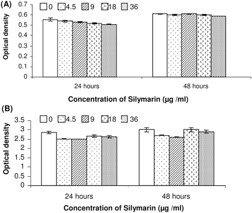
Figure 1.  Effect of silymarin on cellular viability and proliferation of human fibroblast cells. Cells were treated with different concentrations of silymarin for 24 and 48 h. (A) Cell viability was determined by the MTT assay. (B) Cell proliferation was determined by DNA (BrdU) incorporation assay. The data are expressed as mean ± SE from at least three separate experiments.

Figure 2.  Effect of silymarin on hydrogen peroxide-induced damage in fibroblasts using different treatment regimens. (A) Pretreatment only. Fibroblasts were incubated with various concentrations of silymarin overnight before exposure to hydrogen peroxide without silymarin for 3 h. (B) Fibroblasts were pretreated with silymarin overnight and then exposed to hydrogen peroxide and silymarin for 3 h. (C) Fibroblasts were incubated with silymarin and hydrogen peroxide simultaneously for 3 h. The data are expressed as mean ± SE of three separate experiments. Treated cells were compared with cells exposed only to hydrogen peroxide. *p < 0.0.05, **p < 0.01, ***p < 0.001.
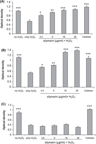
Figure 2.  Effect of silymarin on hydrogen peroxide-induced damage in fibroblasts using different treatment regimens. (A) Pretreatment only. Fibroblasts were incubated with various concentrations of silymarin overnight before exposure to hydrogen peroxide without silymarin for 3 h. (B) Fibroblasts were pretreated with silymarin overnight and then exposed to hydrogen peroxide and silymarin for 3 h. (C) Fibroblasts were incubated with silymarin and hydrogen peroxide simultaneously for 3 h. The data are expressed as mean ± SE of three separate experiments. Treated cells were compared with cells exposed only to hydrogen peroxide. *p < 0.0.05, **p < 0.01, ***p < 0.001.

Figure 3.  Total antioxidant capacity of cell extracts after 3 and 18 h incubation of the cells with various concentrations of silymarin. Total antioxidant capacity was measured by monitoring of ABTS solution decolorization in supernatants of cell homogenates after incubation with silymarin for various times. Results represent mean ± SE, *p < 0.05 compared to the control group.
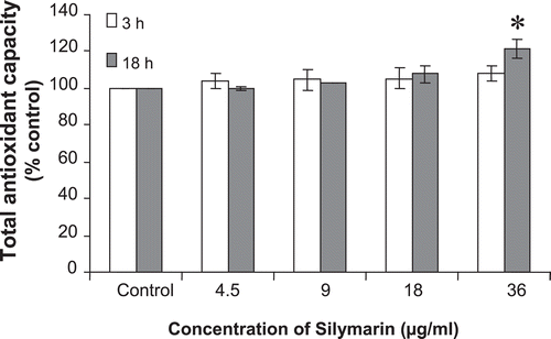
Figure 3.  Total antioxidant capacity of cell extracts after 3 and 18 h incubation of the cells with various concentrations of silymarin. Total antioxidant capacity was measured by monitoring of ABTS solution decolorization in supernatants of cell homogenates after incubation with silymarin for various times. Results represent mean ± SE, *p < 0.05 compared to the control group.

Figure 4.  The effect of silymarin on in vitro collagen synthesis was measured by hydroxyproline assay. Data are present as mean ± SE. No significant difference was seen between groups.
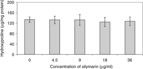
Figure 4.  The effect of silymarin on in vitro collagen synthesis was measured by hydroxyproline assay. Data are present as mean ± SE. No significant difference was seen between groups.

Figure 5.  Real-time RT-PCR measured effect of silymarin on COX-2 mRNA expression of human fibroblast cells. Cells were incubated first with indicated concentrations of silymarin for 18 h and then in the absence or presence of LPS (10 µg/mL) for 24 h. The COX-2 mRNA level was normalized by GAPDH RNA level. Data are mean ± SE of triplicates from an experiment repeated three times. ***p < 0.001 compared with cells treated only with LPS.
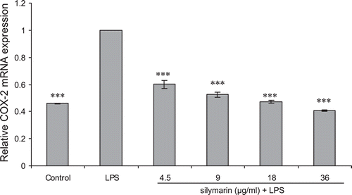
Figure 5.  Real-time RT-PCR measured effect of silymarin on COX-2 mRNA expression of human fibroblast cells. Cells were incubated first with indicated concentrations of silymarin for 18 h and then in the absence or presence of LPS (10 µg/mL) for 24 h. The COX-2 mRNA level was normalized by GAPDH RNA level. Data are mean ± SE of triplicates from an experiment repeated three times. ***p < 0.001 compared with cells treated only with LPS.

Reprints and Corporate Permissions

Please note: Selecting permissions does not provide access to the full text of the article, please see our help page How do I view content?

To request a reprint or corporate permissions for this article, please click on the relevant link below:

Academic Permissions

Please note: Selecting permissions does not provide access to the full text of the article, please see our help page How do I view content?

Obtain permissions instantly via Rightslink by clicking on the button below:

If you are unable to obtain permissions via Rightslink, please complete and submit this Permissions form. For more information, please visit our Permissions help page.