1,374
Views
21
CrossRef citations to date
0
Altmetric
Research Article

Antidiabetic effect of Streblus asper in streptozotocin-induced diabetic rats

, , , &
Pages 369-375 | Received 09 Apr 2012, Accepted 20 Aug 2012, Published online: 14 Feb 2013

Figures & data

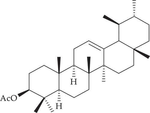
Figure 1.  Structure of α-amyrin acetate.

Figure 1.  Structure of α-amyrin acetate.

Table 1.  Effect of PESA and α-amyrin acetate on glucose levels in STZ-induced diabetic rats.

Table 2.  Effect of PESA and α-amyrin acetate on serum biomarkers in STZ-induced diabetic rats after treatment.

Table 3.  Effect of PESA and α-amyrin acetate on serum lipid profiles in STZ-induced diabetic rats after treatment.

Table 4.  Effect of PESA and α-amyrin acetate on serum insulin, glycosylated hemoglobin (HbA1c) of STZ treated diabetic rats.

Figure 2.  Photomicrographs of 15 day experimental rat liver. Section A (10×) shows a normal histological section with well arranged cells and clear large central vein, cytoplasm and nucleus are well preserved. Section B (10×) shows the complete destruction of hepatocytes degeneration of central vein, fatty degeneration, loss of cell structure and damage in cell membrane in STZ-induced diabetic rat liver. Sections C, D, and E (10×) are treated with PESA (100, 250 & 500 mg/kg) and shows slight necrosis and restored to normal, respectively. Picture F, G & H (10×) depicts no damage and the hepatocyte architecture is near to normal by treatment with α-amyrin acetate (25, 50 & 75 mg/kg). These depict the well preserved hepatocytes, clear cytoplasm and nucleus with less neutrophil infiltration. In case of standard glibenclamide (0.5 mg/kg), I (10×) treatment shows no damage in the hepatocytes and well arranged with central vein.
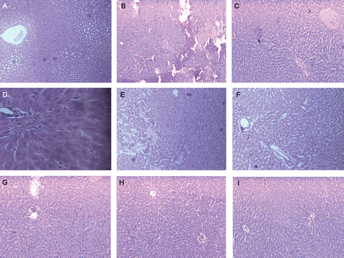
Figure 2.  Photomicrographs of 15 day experimental rat liver. Section A (10×) shows a normal histological section with well arranged cells and clear large central vein, cytoplasm and nucleus are well preserved. Section B (10×) shows the complete destruction of hepatocytes degeneration of central vein, fatty degeneration, loss of cell structure and damage in cell membrane in STZ-induced diabetic rat liver. Sections C, D, and E (10×) are treated with PESA (100, 250 & 500 mg/kg) and shows slight necrosis and restored to normal, respectively. Picture F, G & H (10×) depicts no damage and the hepatocyte architecture is near to normal by treatment with α-amyrin acetate (25, 50 & 75 mg/kg). These depict the well preserved hepatocytes, clear cytoplasm and nucleus with less neutrophil infiltration. In case of standard glibenclamide (0.5 mg/kg), I (10×) treatment shows no damage in the hepatocytes and well arranged with central vein.

Reprints and Corporate Permissions

Please note: Selecting permissions does not provide access to the full text of the article, please see our help page How do I view content?

To request a reprint or corporate permissions for this article, please click on the relevant link below:

Academic Permissions

Please note: Selecting permissions does not provide access to the full text of the article, please see our help page How do I view content?

Obtain permissions instantly via Rightslink by clicking on the button below:

If you are unable to obtain permissions via Rightslink, please complete and submit this Permissions form. For more information, please visit our Permissions help page.