Figures & data
Figure 1. Effects of EFC on paw swelling and polyarthritis index in an AA rat model [n = 6, mean (S.D.)]. Noninjected hind paw swelling (A) and polyarthritis index (B) were assessed at each point at the respective time.
![Figure 1. Effects of EFC on paw swelling and polyarthritis index in an AA rat model [n = 6, mean (S.D.)]. Noninjected hind paw swelling (A) and polyarthritis index (B) were assessed at each point at the respective time.](/cms/asset/6c2b8409-275e-42e7-8925-aad09fca63c3/iphb_a_766892_f0001_b.jpg)
Figure 2. Effects of EFC on body weights and index of immune organs of AA rats [n = 6, mean (S.D.)]. Changes in body weights (A) and index of organs (B) were assessed at each point at the respective time. *p < 0.05, **p < 0.01 versus the model group.
![Figure 2. Effects of EFC on body weights and index of immune organs of AA rats [n = 6, mean (S.D.)]. Changes in body weights (A) and index of organs (B) were assessed at each point at the respective time. *p < 0.05, **p < 0.01 versus the model group.](/cms/asset/795e3d13-c1b8-48a0-b536-ceae00ff9a02/iphb_a_766892_f0002_b.jpg)
Figure 3. Histopathological examination of ankle joints stained with hematoxylin and eosin under a light microscope: histological features with cartilage erosion (A), cellular infiltration (B) and pannus formation (C) from six groups.
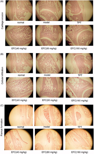
Figure 4. Histopathological evaluation of ankle joint in AA rat models. Histological appearance was scored for the presence of synovial proliferation, cellular infiltration, cartilage erosion and pannus formation. *p < 0.05, **p < 0.01 compared with the model group [n = 6, mean (S.D.)].
![Figure 4. Histopathological evaluation of ankle joint in AA rat models. Histological appearance was scored for the presence of synovial proliferation, cellular infiltration, cartilage erosion and pannus formation. *p < 0.05, **p < 0.01 compared with the model group [n = 6, mean (S.D.)].](/cms/asset/31ee3cb0-eb43-4a4c-94d1-1cd6b076c041/iphb_a_766892_f0004_b.jpg)
Table 1. Effect of EFC on hemorheological disorders in rats.
Table 2. Effect of EFC on the IL-1 and TNF-α levels in rats serum.