
Figures & data
Figure 1. Microscopical characters of Desmostachya bipinnata leaf. (a) T.S. of leaf lamina, (b) T.S. of leaf margin, bundle sheath cells, (c) vascular bundles, phloem, xylem, sclerenchymatous cells, (d) cuticle and bulliform cells and (e) T.S. of mid rib with lamina.
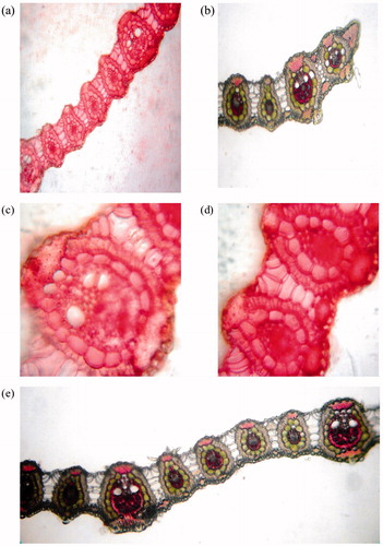
Figure 2. Microscopical characters of Desmostachya bipinnata stem. (a) Starch with oil glands, (b) cortical region, sclerenchymatous sheath followed by vascular bundles, (c) scattered vascular bundles, (d) emerging root from node, (e) meta xylem and pholeum and (f) sclerenchymatous sheath.
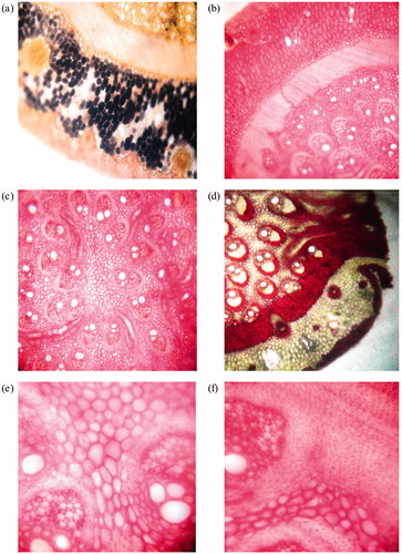
Figure 3. Microscopical characters of Desmostachya bipinnata root. (a) T.S. of root, (b) T.S. showing steler region having sclerenchymatous cells, (c) endodermis, metaxylem and phloem and (d) epidermis, trichomes and aerenchymatous cells.
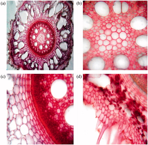
Table 1. Microchemical tests performed with various plant parts of Desmostachya bipinnata (L.) Stapf.
Figure 4. Powder characteristics of aerial parts of Desmostachya bipinnata. (a) Lignified vessel, (b) phloem, (c) fiber, parenchymatous cells and (d) fibers, epidermal cells, parenchymatous cells and vessels.
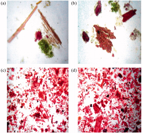
Figure 5. Powder characteristics of underground parts of Desmostachya bipinnata. (a) Endodermal cells, trichomes, (b) sclerenchymatous cells, (c) fiber and (d) starch grains, vessels.
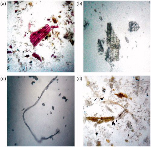
Table 2. Microscopic measurements for several powder characteristics in Desmostachya bipinnata (L.) Stapf.
Table 3. Fluorescence studies with different plant parts of Desmostachya bipinnata (L.) Stapf.
Table 4. Estimated standardization parameters of Desmostachya bipinnata (L.) Stapf as per WHO guidelines.
Table 5. Extractive yields estimated for various plant parts of Desmostachya bipinnata (L.) Stapf.
Table 6. Phytochemical tests performed with various plant parts of Desmostachya bipinnata (L.) Stapf.
Table 7. Chromatographic profile of various plant parts of Desmostachya bipinnata.