
Figures & data
Table 1. Comparison on the phytochemical constituents between the dried leaves of chloroform extracts of M. calabura (MEMC) and M. malabathricum with their respective extract, MEMC or (MEMM).

Table 2. The antioxidant profiles and TPC values of CEMC and CEMM, at the concentration of 200 μg/ml, as assessed using the respective oxidative assays.
Table 3. Effects of CEMC or CEMM on ethanol-induced gastric ulcer model in rats.
Figure 2. Macroscopic analysis of the rat stomach pre-treated with the respective test solution followed by the ethanol-induced gastric ulcer formation. (A) Normal untreated stomach shows normal surface appearance and absence of ulcer formation. (B) Pretreatment with vehicle (8% Tween 80; negative control) shows severe hemorrhagic ulcer and lesions of the surface of the stomach. (C) Pretreatment with 100 mg/kg ranitidine (positive control) shows almost normal surface appearance with slight gastric ulcer formation indicated by reduced in hemorrhagic tissue appearance. (D1) Pretreatment with 50 mg/kg CEMC shows severe gastric ulcer formation and marked hemorrhagic streaks appearance. (D2) Pretreatment with 250 mg/kg CEMC shows mild gastric ulceration and reduction in hemorrhagic streaks appearance. (D3) Pretreatment with 500 mg/kg CEMC shows complete absence of ulcer formation and lack of hemorrhagic streaks appearance. (E1) Pretreatment with 50 mg/kg CEMM shows severe ulceration and marked hemorrhagic streaks appearance. (E2) Pretreatment with 250 mg/kg CEMM shows mild gastric ulcer formation and marked reduction in hemorrhagic streaks appearance. (E3) Pretreatment with 500 mg/kg CEMM shows complete absence of gastric ulcer formation and marked decrease in hemorrhagic streaks appearance.
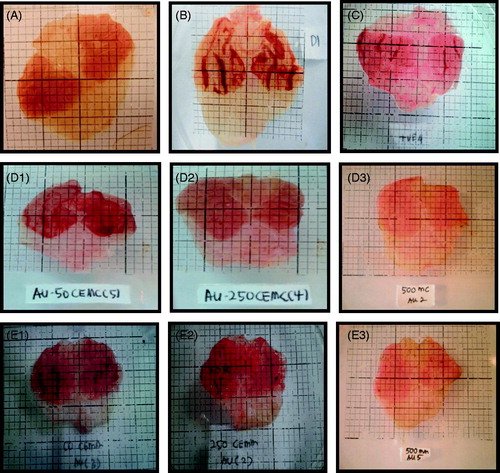
Figure 3. (A) Microscopic analysis at the ×10 magnification of the rat stomach pre-treated with the respective test solution followed by the ethanol-induced gastric ulcer formation. (i) Gastric tissue of normal (untreated) group shows mucosa layer (M) consist of gastric glands, which extend from the level of muscularis mucosa (MM) to open into the stomach lumen via gastric pits (GP). (ii) Gastric tissue of vehicle (negative control)-treated group shows a total disruption of the epithelial layer (EL) of the mucosa and GP. (iii) Gastric tissue of 100 mg/kg ranitidine (positive control)-treated group shows the irregular architecture of the epithelial surface of mucosa, which is still in the recovery phase. (iv) Gastric tissue of 50 mg/kg CEMC-treated group shows the total disruption of EL indicated by the slough off of the mucosa lining that had occurred in a few places. Structure of GP was also disrupted and the mucosa layer show presence of inflammatory cells. (v) Gastric tissue of 250 mg/kg CEMC-treated group shows that the EL is undergoing recovery indicated by the tissue architecture that resemble the normal lining of stomach. The cells in the mucosa were not disrupted as the lesions on the surface are fewer. (vi) Gastric tissue of 500 mg/kg CEMC-treated group shows that the EL lining the stomach was almost in its normal form and lesions are hardly found at the mucosa layer. (vii) Gastric tissue of 50 mg/kg CEMM-treated group shows mild disruption of the EL with the slough off of the surface of the mucosa. Edema is present at the MM layer. (viii) Gastric tissue of 250 mg/kg CEMM-treated group shows that the mucosa layer is highly populated with H and N. Morphology of the cells were also changed and did not resemble normal structure of cells. (ix) Gastric tissue of 500 mg/kg CEMM-treated group shows improvement in the structure of the mucosa with the morphology of the cells resembles those of the normal untreated cells. The surface of EL shows better lining at the top of mucosa layer (Hematoxylin & eosin; magnification ×10). (B) Microscopic analysis at the ×40 magnification of the rat’s stomach pre-treated with the respective test solution followed by the ethanol-induced gastric ulcer formation. (i) Gastric tissue of normal (untreated) group shows that the gastric glands contain mixed population of cells such as parietal cell (P), which is acid-secreting cells and chief cells (C) that are located towards the bases of the gastric glands. Mucus will line the GP in which gastric gland will open. (ii) Gastric tissue of vehicle (negative control)-treated group shows a high number of neutrophils (N) and hemorrhage (H) within the mucosa layer. (iii) Gastric tissue of 100 mg/kg ranitidine (positive control)-treated group shows the presence of inflammatory exudates mainly N and mild H within the mucosa layer. (iv) Gastric tissue of 50 mg/kg CEMC-treated group shows the presence of H and N, which can be seen clearly and mostly reside at the base of the mucosa layer. (v) Gastric tissue of 250 mg/kg CEMC-treated group also shows that N were present at the base of the mucosa layer with the absence of H. (vi) Gastric tissue of 500 mg/kg CEMC-treated group shows normal structure of the P and C cells with the absence of H and inflammatory exudates. (vii) Gastric tissue of 50 mg/kg CEMM-treated group shows the presence of H and N, which were found mainly at the base of the mucosa layer. (viii) Gastric tissue of 250 mg/kg CEMM-treated group shows the presence of low number of N and H that were mainly located at the base of mucosa layer. (ix) Gastric tissue of 500 mg/kg CEMM-treated group shows the least number of N and H, and the lining of the mucosa had almost the same architecture as the normal stomach (hematoxylin & eosin; magnification ×40).
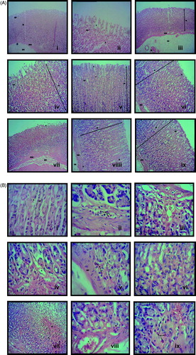
Table 4. Effects of CEMC or CEMM on pylorus ligation-induced gastric ulcer model in rats.